 Movie
Movie Controller

[English] 日本語
 Yorodumi
- PDB-7wbo: Crystal structure of Sarcoplasmic Calcium-Binding Protein from Sc... -
+ Open data

- Basic information
| Entry | Database: PDB / ID: 7wbo | ||||||
|---|---|---|---|---|---|---|---|
| Title | Crystal structure of Sarcoplasmic Calcium-Binding Protein from Scylla paramamosain | ||||||
 Components Components | Sarcoplasmic calcium-binding protein | ||||||
 Keywords Keywords | ALLERGEN / allergy / Scylla paramamosain / EF-hand | ||||||
| Function / homology |  Function and homology information Function and homology information | ||||||
| Biological species |  Scylla paramamosain (green mud crab) Scylla paramamosain (green mud crab) | ||||||
| Method |  X-RAY DIFFRACTION / SYNCHROTRON / MOLECULAR REPLACEMENT / molecular replacement / Resolution: 1.6 Å X-RAY DIFFRACTION / SYNCHROTRON / MOLECULAR REPLACEMENT / molecular replacement / Resolution: 1.6 Å | ||||||
 Authors Authors | Chen, Y. / Jin, T. / Liu, G. | ||||||
| Funding support | 1items
| ||||||
 Citation Citation | Journal: J.Agric.Food Chem. / Year: 2023 Title: Crystal Structure Analysis of Sarcoplasmic-Calcium-Binding Protein: An Allergen in Scylla paramamosain. Authors: Chen, Y. / Jin, T. / Li, M. / Yun, X. / Huan, F. / Liu, Q. / Hu, M. / Wei, X. / Zheng, P. / Liu, G. | ||||||
| History |
|
- Structure visualization
| Structure viewer | Molecule: MolmilJmol/JSmol |
|---|
- Downloads & links
-Download
| PDBx/mmCIF format | 7wbo.cif.gz | 63 KB | Display | PDBx/mmCIF format |
|---|---|---|---|---|
| PDB format | pdb7wbo.ent.gz | 41.9 KB | Display | PDB format |
| PDBx/mmJSON format | 7wbo.json.gz | Tree view | PDBx/mmJSON format | |
| Others |  Other downloads Other downloads |
-Validation report
| Arichive directory | https://data.pdbj.org/pub/pdb/validation_reports/wb/7wboftp://data.pdbj.org/pub/pdb/validation_reports/wb/7wbo | HTTPS FTP |
|---|
-Related structure data
| Related structure data |  3akaS S: Starting model for refinement |
|---|---|
| Similar structure data |
-Links
 PDBj
PDBj- Assembly
| Deposited unit | 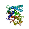
| ||||||||
|---|---|---|---|---|---|---|---|---|---|
| 1 |
| ||||||||
| Unit cell |
|
-Components
| #1: Protein | Mass: 22360.053 Da / Num. of mol.: 1 Source method: isolated from a genetically manipulated source Source: (gene. exp.) Scylla paramamosain (green mud crab) / Production host:  | ||||||||
|---|---|---|---|---|---|---|---|---|---|
| #2: Chemical | ChemComp-EDO /   Mass: 62.068 Da / Num. of mol.: 7 / Source method: obtained synthetically / Formula: C2H6O2 Mass: 62.068 Da / Num. of mol.: 7 / Source method: obtained synthetically / Formula: C2H6O2#3: Chemical | ChemComp-CA /   Mass: 40.078 Da / Num. of mol.: 4 / Source method: obtained synthetically / Formula: Ca / Feature type: SUBJECT OF INVESTIGATION Mass: 40.078 Da / Num. of mol.: 4 / Source method: obtained synthetically / Formula: Ca / Feature type: SUBJECT OF INVESTIGATION#4: Chemical | ChemComp-NA / |   Mass: 22.990 Da / Num. of mol.: 1 / Source method: obtained synthetically / Formula: Na Mass: 22.990 Da / Num. of mol.: 1 / Source method: obtained synthetically / Formula: Na#5: Water | ChemComp-HOH / |  Mass: 18.015 Da / Num. of mol.: 250 / Source method: isolated from a natural source / Formula: H2O Mass: 18.015 Da / Num. of mol.: 250 / Source method: isolated from a natural source / Formula: H2OHas ligand of interest | Y | |
-Experimental details
-Experiment
| Experiment | Method: X-RAY DIFFRACTION / Number of used crystals: 1 |
|---|
- Sample preparation
| Crystal | Density Matthews: 2.27 Å3/Da / Density % sol: 45.93 % |
|---|---|
| Crystal grow | Temperature: 298 K / Method: vapor diffusion, hanging drop / Details: 20% (v/v) PEG 3350, 0.2M NH4Cl |
-Data collection
| Diffraction | Mean temperature: 100 K / Serial crystal experiment: N |
|---|---|
| Diffraction source | Source: SYNCHROTRON / Site: NFPSS  / Beamline: BL19U1 / Wavelength: 0.97852 Å / Beamline: BL19U1 / Wavelength: 0.97852 Å |
| Detector | Type: DECTRIS PILATUS3 6M / Detector: PIXEL / Date: Dec 15, 2019 |
| Radiation | Protocol: SINGLE WAVELENGTH / Monochromatic (M) / Laue (L): M / Scattering type: x-ray |
| Radiation wavelength | Wavelength: 0.97852 Å / Relative weight: 1 |
| Reflection | Resolution: 1.6→50 Å / Num. obs: 24946 / % possible obs: 95.5 % / Redundancy: 3.7 % / CC1/2: 0.98 / Rmerge(I) obs: 0.055 / Rrim(I) all: 0.033 / Net I/σ(I): 18.6 |
| Reflection shell | Resolution: 1.6→1.63 Å / Redundancy: 3.8 % / Rmerge(I) obs: 0.082 / Mean I/σ(I) obs: 15 / Num. unique obs: 1235 / CC1/2: 0.986 / Rpim(I) all: 0.048 / % possible all: 95.4 |
-Phasing
| Phasing | Method: molecular replacement | ||||||
|---|---|---|---|---|---|---|---|
| Phasing MR | R rigid body: 0.605
|
- Processing
| Software |
| ||||||||||||||||||||||||||||||||||||||||||||||||||||||||||||
|---|---|---|---|---|---|---|---|---|---|---|---|---|---|---|---|---|---|---|---|---|---|---|---|---|---|---|---|---|---|---|---|---|---|---|---|---|---|---|---|---|---|---|---|---|---|---|---|---|---|---|---|---|---|---|---|---|---|---|---|---|---|
| Refinement | Method to determine structure: MOLECULAR REPLACEMENT Starting model: 3AKA Resolution: 1.6→40.52 Å / Cor.coef. Fo:Fc: 0.964 / Cor.coef. Fo:Fc free: 0.938 / WRfactor Rfree: 0.2156 / WRfactor Rwork: 0.175 / FOM work R set: 0.8991 / SU B: 1.246 / SU ML: 0.046 / SU R Cruickshank DPI: 0.0818 / SU Rfree: 0.085 / Cross valid method: THROUGHOUT / σ(F): 0 / ESU R: 0.082 / ESU R Free: 0.085 / Stereochemistry target values: MAXIMUM LIKELIHOOD Details: HYDROGENS HAVE BEEN ADDED IN THE RIDING POSITIONS U VALUES : REFINED INDIVIDUALLY
| ||||||||||||||||||||||||||||||||||||||||||||||||||||||||||||
| Solvent computation | Ion probe radii: 0.8 Å / Shrinkage radii: 0.8 Å / VDW probe radii: 1.2 Å / Solvent model: MASK | ||||||||||||||||||||||||||||||||||||||||||||||||||||||||||||
| Displacement parameters | Biso max: 56.7 Å2 / Biso mean: 14.713 Å2 / Biso min: 5.03 Å2
| ||||||||||||||||||||||||||||||||||||||||||||||||||||||||||||
| Refinement step | Cycle: final / Resolution: 1.6→40.52 Å
| ||||||||||||||||||||||||||||||||||||||||||||||||||||||||||||
| Refine LS restraints |
| ||||||||||||||||||||||||||||||||||||||||||||||||||||||||||||
| LS refinement shell | Resolution: 1.601→1.643 Å / Rfactor Rfree error: 0 / Total num. of bins used: 20
|
