 Movie
Movie Controller


+ Open data

- Basic information
| Entry | Database: PDB / ID: 7wbg | ||||||
|---|---|---|---|---|---|---|---|
| Title | BF2*1901/RY8 | ||||||
 Components Components |
| ||||||
 Keywords Keywords | IMMUNE SYSTEM / MHC I | ||||||
| Function / homology |  Function and homology information Function and homology informationER-Phagosome pathway / Endosomal/Vacuolar pathway / Immunoregulatory interactions between a Lymphoid and a non-Lymphoid cell / DAP12 signaling / Antigen Presentation: Folding, assembly and peptide loading of class I MHC / antigen processing and presentation of peptide antigen via MHC class I / Neutrophil degranulation / cellular response to iron ion / peptide antigen assembly with MHC class II protein complex / negative regulation of forebrain neuron differentiation ...ER-Phagosome pathway / Endosomal/Vacuolar pathway / Immunoregulatory interactions between a Lymphoid and a non-Lymphoid cell / DAP12 signaling / Antigen Presentation: Folding, assembly and peptide loading of class I MHC / antigen processing and presentation of peptide antigen via MHC class I / Neutrophil degranulation / cellular response to iron ion / peptide antigen assembly with MHC class II protein complex / negative regulation of forebrain neuron differentiation / MHC class II protein complex / peptide antigen assembly with MHC class I protein complex / regulation of iron ion transport / HFE-transferrin receptor complex / MHC class I peptide loading complex / positive regulation of T cell cytokine production / antigen processing and presentation of endogenous peptide antigen via MHC class I / antigen processing and presentation of exogenous peptide antigen via MHC class II / positive regulation of immune response / MHC class I protein complex / peptide antigen binding / positive regulation of T cell activation / negative regulation of neurogenesis / positive regulation of receptor-mediated endocytosis / cellular response to nicotine / negative regulation of epithelial cell proliferation / MHC class II protein complex binding / late endosome membrane / positive regulation of cellular senescence / protein homotetramerization / amyloid fibril formation / intracellular iron ion homeostasis / learning or memory / immune response / lysosomal membrane / structural molecule activity / Golgi apparatus / protein homodimerization activity / extracellular region / cytosol Similarity search - Function | ||||||
| Biological species |   uncultured virus (environmental samples) uncultured virus (environmental samples) | ||||||
| Method |  X-RAY DIFFRACTION / SYNCHROTRON / MOLECULAR REPLACEMENT / Resolution: 2 Å X-RAY DIFFRACTION / SYNCHROTRON / MOLECULAR REPLACEMENT / Resolution: 2 Å | ||||||
 Authors Authors | Liu, W.J. | ||||||
| Funding support |  China, 1items China, 1items
| ||||||
 Citation Citation | Journal: J Immunol. / Year: 2023 Title: A Wider and Deeper Peptide-Binding Groove for the Class I Molecules from B15 Compared with B19 Chickens Correlates with Relative Resistance to Marek's Disease. Authors: Han, L. / Wu, S. / Zhang, T. / Peng, W. / Zhao, M. / Yue, C. / Wen, W. / Cai, W. / Li, M. / Wallny, H.J. / Avila, D.W. / Mwangi, W. / Nair, V. / Ternette, N. / Guo, Y. / Zhao, Y. / Chai, Y. ...Authors: Han, L. / Wu, S. / Zhang, T. / Peng, W. / Zhao, M. / Yue, C. / Wen, W. / Cai, W. / Li, M. / Wallny, H.J. / Avila, D.W. / Mwangi, W. / Nair, V. / Ternette, N. / Guo, Y. / Zhao, Y. / Chai, Y. / Qi, J. / Liang, H. / Gao, G.F. / Kaufman, J. / Liu, W.J. | ||||||
| History |
|
- Structure visualization
| Structure viewer | Molecule: MolmilJmol/JSmol |
|---|
- Downloads & links
-Download
| PDBx/mmCIF format | 7wbg.cif.gz | 211.5 KB | Display | PDBx/mmCIF format |
|---|---|---|---|---|
| PDB format | pdb7wbg.ent.gz | 135.2 KB | Display | PDB format |
| PDBx/mmJSON format | 7wbg.json.gz | Tree view | PDBx/mmJSON format | |
| Others |  Other downloads Other downloads |
-Validation report
| Arichive directory | https://data.pdbj.org/pub/pdb/validation_reports/wb/7wbgftp://data.pdbj.org/pub/pdb/validation_reports/wb/7wbg | HTTPS FTP |
|---|
-Related structure data
| Related structure data | 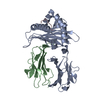 7wbiC  2yezS S: Starting model for refinement C: citing same article ( |
|---|---|
| Similar structure data |
-Links
 PDBj
PDBj- Assembly
| Deposited unit | 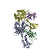
| ||||||||||||
|---|---|---|---|---|---|---|---|---|---|---|---|---|---|
| 1 | 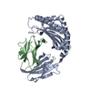
| ||||||||||||
| 2 | 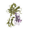
| ||||||||||||
| Unit cell |
|
-Components
| #1: Protein | Mass: 30801.967 Da / Num. of mol.: 2 Source method: isolated from a genetically manipulated source Source: (gene. exp.)  #2: Protein | Mass: 11062.404 Da / Num. of mol.: 2 Source method: isolated from a genetically manipulated source Source: (gene. exp.) #3: Protein/peptide | Mass: 1126.205 Da / Num. of mol.: 2 / Source method: obtained synthetically / Source: (synth.) uncultured virus (environmental samples)#4: Water | ChemComp-HOH / |  Mass: 18.015 Da / Num. of mol.: 589 / Source method: isolated from a natural source / Formula: H2O Mass: 18.015 Da / Num. of mol.: 589 / Source method: isolated from a natural source / Formula: H2OHas protein modification | Y | |
|---|
-Experimental details
-Experiment
| Experiment | Method: X-RAY DIFFRACTION / Number of used crystals: 1 |
|---|
- Sample preparation
| Crystal | Density Matthews: 2.14 Å3/Da / Density % sol: 42.54 % |
|---|---|
| Crystal grow | Temperature: 291.15 K / Method: vapor diffusion, sitting drop Details: 0.15 M Potassium bromide and 30% w/v Polyethylene glycol monomethyl ether 2,000 |
-Data collection
| Diffraction | Mean temperature: 100 K / Serial crystal experiment: N |
|---|---|
| Diffraction source | Source: SYNCHROTRON / Site: SSRF / Beamline: BL17U1 / Wavelength: 0.97853 Å |
| Detector | Type: DECTRIS PILATUS 6M / Detector: PIXEL / Date: Jan 14, 2014 |
| Radiation | Protocol: SINGLE WAVELENGTH / Monochromatic (M) / Laue (L): M / Scattering type: x-ray |
| Radiation wavelength | Wavelength: 0.97853 Å / Relative weight: 1 |
| Reflection | Resolution: 2→50 Å / Num. obs: 49775 / % possible obs: 98.2 % / Redundancy: 6.3 % / Biso Wilson estimate: 23.42 Å2 / Rmerge(I) obs: 0.068 / Net I/σ(I): 13.1 |
| Reflection shell | Resolution: 2→2.07 Å / Rmerge(I) obs: 0.142 / Num. unique obs: 4913 |
- Processing
| Software |
| |||||||||||||||||||||||||||||||||||||||||||||||||||||||||||||||||||||||||||||||||||||||||||||||||||||||||||||||||||||||||||||||||||||
|---|---|---|---|---|---|---|---|---|---|---|---|---|---|---|---|---|---|---|---|---|---|---|---|---|---|---|---|---|---|---|---|---|---|---|---|---|---|---|---|---|---|---|---|---|---|---|---|---|---|---|---|---|---|---|---|---|---|---|---|---|---|---|---|---|---|---|---|---|---|---|---|---|---|---|---|---|---|---|---|---|---|---|---|---|---|---|---|---|---|---|---|---|---|---|---|---|---|---|---|---|---|---|---|---|---|---|---|---|---|---|---|---|---|---|---|---|---|---|---|---|---|---|---|---|---|---|---|---|---|---|---|---|---|---|
| Refinement | Method to determine structure: MOLECULAR REPLACEMENT Starting model: 2YEZ Resolution: 2→33.4 Å / SU ML: 0.202 / Cross valid method: NONE / σ(F): 1.34 / Phase error: 21.7077 / Stereochemistry target values: GeoStd + Monomer Library
| |||||||||||||||||||||||||||||||||||||||||||||||||||||||||||||||||||||||||||||||||||||||||||||||||||||||||||||||||||||||||||||||||||||
| Solvent computation | Shrinkage radii: 0.9 Å / VDW probe radii: 1.11 Å / Solvent model: FLAT BULK SOLVENT MODEL | |||||||||||||||||||||||||||||||||||||||||||||||||||||||||||||||||||||||||||||||||||||||||||||||||||||||||||||||||||||||||||||||||||||
| Displacement parameters | Biso mean: 27.36 Å2 | |||||||||||||||||||||||||||||||||||||||||||||||||||||||||||||||||||||||||||||||||||||||||||||||||||||||||||||||||||||||||||||||||||||
| Refinement step | Cycle: LAST / Resolution: 2→33.4 Å
| |||||||||||||||||||||||||||||||||||||||||||||||||||||||||||||||||||||||||||||||||||||||||||||||||||||||||||||||||||||||||||||||||||||
| Refine LS restraints |
| |||||||||||||||||||||||||||||||||||||||||||||||||||||||||||||||||||||||||||||||||||||||||||||||||||||||||||||||||||||||||||||||||||||
| LS refinement shell |
|
