 Movie
Movie Controller


+ Open data

- Basic information
| Entry | Database: PDB / ID: 7wbb | |||||||||||||||||||||
|---|---|---|---|---|---|---|---|---|---|---|---|---|---|---|---|---|---|---|---|---|---|---|
| Title | Cryo-EM structure of substrate engaged Drg1 hexamer | |||||||||||||||||||||
 Components Components |
| |||||||||||||||||||||
 Keywords Keywords | ATP-binding protein / cryo-EM structure of Drg1 / RIBOSOME | |||||||||||||||||||||
| Function / homology |  Function and homology information Function and homology informationmitotic spindle disassembly / VCP-NPL4-UFD1 AAA ATPase complex / retrograde protein transport, ER to cytosol / polyubiquitin modification-dependent protein binding / autophagosome maturation / ATP hydrolysis activity / ATP binding / nucleus / cytosol Similarity search - Function | |||||||||||||||||||||
| Biological species |   | |||||||||||||||||||||
| Method | ELECTRON MICROSCOPY / single particle reconstruction / cryo EM / Resolution: 3.6 Å | |||||||||||||||||||||
 Authors Authors | Ma, C.Y. / Wu, D.M. / Chen, Q. / Gao, N. | |||||||||||||||||||||
| Funding support | 1items
| |||||||||||||||||||||
 Citation Citation | Journal: Nat Commun / Year: 2022 Title: Structural dynamics of AAA + ATPase Drg1 and mechanism of benzo-diazaborine inhibition. Authors: Chengying Ma / Damu Wu / Qian Chen / Ning Gao /  Abstract: The type II AAA + ATPase Drg1 is a ribosome assembly factor, functioning to release Rlp24 from the pre-60S particle just exported from nucleus, and its activity in can be inhibited by a drug ...The type II AAA + ATPase Drg1 is a ribosome assembly factor, functioning to release Rlp24 from the pre-60S particle just exported from nucleus, and its activity in can be inhibited by a drug molecule diazaborine. However, molecular mechanisms of Drg1-mediated Rlp24 removal and diazaborine-mediated inhibition are not fully understood. Here, we report Drg1 structures in different nucleotide-binding and benzo-diazaborine treated states. Drg1 hexamers transits between two extreme conformations (planar or helical arrangement of protomers). By forming covalent adducts with ATP molecules in both ATPase domain, benzo-diazaborine locks Drg1 hexamers in a symmetric and non-productive conformation to inhibits both inter-protomer and inter-ring communication of Drg1 hexamers. We also obtained a substrate-engaged mutant Drg1 structure, in which conserved pore-loops form a spiral staircase to interact with the polypeptide through a sequence-independent manner. Structure-based mutagenesis data highlight the functional importance of the pore-loop, the D1-D2 linker and the inter-subunit signaling motif of Drg1, which share similar regulatory mechanisms with p97. Our results suggest that Drg1 may function as an unfoldase that threads a substrate protein within the pre-60S particle. | |||||||||||||||||||||
| History |
|
- Structure visualization
| Structure viewer | Molecule: MolmilJmol/JSmol |
|---|
- Downloads & links
-Download
| PDBx/mmCIF format | 7wbb.cif.gz | 715 KB | Display | PDBx/mmCIF format |
|---|---|---|---|---|
| PDB format | pdb7wbb.ent.gz | 600.9 KB | Display | PDB format |
| PDBx/mmJSON format | 7wbb.json.gz | Tree view | PDBx/mmJSON format | |
| Others |  Other downloads Other downloads |
-Validation report
| Summary document | 7wbb_validation.pdf.gz | 1.5 MB | Display | wwPDB validaton report |
|---|---|---|---|---|
| Full document | 7wbb_full_validation.pdf.gz | 1.6 MB | Display | |
| Data in XML | 7wbb_validation.xml.gz | 111.9 KB | Display | |
| Data in CIF | 7wbb_validation.cif.gz | 169.7 KB | Display | |
| Arichive directory | https://data.pdbj.org/pub/pdb/validation_reports/wb/7wbbftp://data.pdbj.org/pub/pdb/validation_reports/wb/7wbb | HTTPS FTP |
-Related structure data
| Related structure data |  32396MC  7wd3C 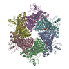 7ykkC  7yklC  7yktC 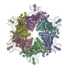 7ykzC M: map data used to model this data C: citing same article ( |
|---|---|
| Similar structure data |

-Links
 PDBj
PDBj


- Assembly
| Deposited unit | 
|
|---|---|
| 1 |
|
-Components
| #1: Protein | Mass: 84848.742 Da / Num. of mol.: 6 / Mutation: E346Q, E617Q Source method: isolated from a genetically manipulated source Source: (gene. exp.) Gene: AFG2 / Production host: #2: Protein/peptide | | Mass: 1975.426 Da / Num. of mol.: 1 Source method: isolated from a genetically manipulated source Source: (gene. exp.) #3: Chemical | ChemComp-ATP /   Mass: 507.181 Da / Num. of mol.: 11 / Source method: obtained synthetically / Formula: C10H16N5O13P3 / Feature type: SUBJECT OF INVESTIGATION / Comment: ATP, energy-carrying molecule*YM Mass: 507.181 Da / Num. of mol.: 11 / Source method: obtained synthetically / Formula: C10H16N5O13P3 / Feature type: SUBJECT OF INVESTIGATION / Comment: ATP, energy-carrying molecule*YMHas ligand of interest | Y | Has protein modification | N | |
|---|
-Experimental details
-Experiment
| Experiment | Method: ELECTRON MICROSCOPY |
|---|---|
| EM experiment | Aggregation state: PARTICLE / 3D reconstruction method: single particle reconstruction |
- Sample preparation
| Component | Name: Double mutant Drg1 in the presence of ATP with substrate engaged Type: COMPLEX / Entity ID: #1-#2 / Source: RECOMBINANT |
|---|---|
| Source (natural) | Organism: |
| Source (recombinant) | Organism: |
| Buffer solution | pH: 8 |
| Specimen | Embedding applied: NO / Shadowing applied: NO / Staining applied: NO / Vitrification applied: YES |
| Specimen support | Grid type: Quantifoil R1.2/1.3 |
| Vitrification | Cryogen name: ETHANE / Humidity: 100 % |
- Electron microscopy imaging
| Experimental equipment |  Model: Titan Krios / Image courtesy: FEI Company |
|---|---|
| Microscopy | Model: FEI TITAN KRIOS |
| Electron gun | Electron source:  FIELD EMISSION GUN / Accelerating voltage: 300 kV / Illumination mode: FLOOD BEAM FIELD EMISSION GUN / Accelerating voltage: 300 kV / Illumination mode: FLOOD BEAM |
| Electron lens | Mode: BRIGHT FIELD / Nominal defocus max: 2500 nm / Nominal defocus min: 1500 nm |
| Image recording | Electron dose: 8 e/Å2 / Film or detector model: GATAN K2 QUANTUM (4k x 4k) |
- Processing
| Software | Name: PHENIX / Version: 1.19.2_4158: / Classification: refinement | ||||||||||||||||||||||||
|---|---|---|---|---|---|---|---|---|---|---|---|---|---|---|---|---|---|---|---|---|---|---|---|---|---|
| EM software | Name: PHENIX / Category: model refinement | ||||||||||||||||||||||||
| CTF correction | Type: NONE | ||||||||||||||||||||||||
| 3D reconstruction | Resolution: 3.6 Å / Resolution method: FSC 0.143 CUT-OFF / Num. of particles: 152973 / Symmetry type: POINT | ||||||||||||||||||||||||
| Refine LS restraints |
|
