 Movie
Movie Controller


+ Open data

- Basic information
| Entry | Database: PDB / ID: 7wa9 | |||||||||||||||
|---|---|---|---|---|---|---|---|---|---|---|---|---|---|---|---|---|
| Title | Crystal structure of MSMEG_5634 from Mycobacterium smegmatis | |||||||||||||||
 Components Components | MSMEG_5634 | |||||||||||||||
 Keywords Keywords | LIPID BINDING PROTEIN / MSMEG_5634 / acyl-AcpM / FAS-II inhibitor / Mycobacterium smegmatis | |||||||||||||||
| Function / homology | Polyketide cyclase/dehydrase / Polyketide cyclase / dehydrase and lipid transport / START domain / Alpha-D-Glucose-1,6-Bisphosphate; Chain A, domain 4 / START-like domain superfamily / 2-Layer Sandwich / Alpha Beta / Toxin Function and homology information Function and homology information | |||||||||||||||
| Biological species |  Mycolicibacterium smegmatis MC2 155 (bacteria) Mycolicibacterium smegmatis MC2 155 (bacteria) | |||||||||||||||
| Method |  X-RAY DIFFRACTION / SYNCHROTRON / SAD / Resolution: 1.9 Å X-RAY DIFFRACTION / SYNCHROTRON / SAD / Resolution: 1.9 Å | |||||||||||||||
 Authors Authors | Wang, Z. / Zhang, W. | |||||||||||||||
| Funding support |  China, 4items China, 4items
| |||||||||||||||
 Citation Citation | Journal: Front Microbiol / Year: 2022 Title: A Novel Acyl-AcpM-Binding Protein Confers Intrinsic Sensitivity to Fatty Acid Synthase Type II Inhibitors in Mycobacterium smegmatis Authors: Li, M. / Huang, Q. / Zhang, W. / Cao, Y. / Wang, Z. / Zhao, Z. / Zhang, X. / Zhang, J. | |||||||||||||||
| History |
|
- Structure visualization
| Structure viewer | Molecule: MolmilJmol/JSmol |
|---|
- Downloads & links
-Download
| PDBx/mmCIF format | 7wa9.cif.gz | 44.4 KB | Display | PDBx/mmCIF format |
|---|---|---|---|---|
| PDB format | pdb7wa9.ent.gz | 28.5 KB | Display | PDB format |
| PDBx/mmJSON format | 7wa9.json.gz | Tree view | PDBx/mmJSON format | |
| Others |  Other downloads Other downloads |
-Validation report
| Arichive directory | https://data.pdbj.org/pub/pdb/validation_reports/wa/7wa9ftp://data.pdbj.org/pub/pdb/validation_reports/wa/7wa9 | HTTPS FTP |
|---|
-Related structure data
| Similar structure data |
|---|
-Links
 PDBj
PDBj- Assembly
| Deposited unit | 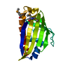
| ||||||||||||
|---|---|---|---|---|---|---|---|---|---|---|---|---|---|
| 1 |
| ||||||||||||
| Unit cell |
|
-Components
| #1: Protein | Mass: 16407.494 Da / Num. of mol.: 1 Source method: isolated from a genetically manipulated source Source: (gene. exp.) Mycolicibacterium smegmatis MC2 155 (bacteria)Strain: ATCC 700084 / mc(2)155 / Gene: MSMEG_5634 / Production host: |
|---|---|
| #2: Water | ChemComp-HOH /  Mass: 18.015 Da / Num. of mol.: 52 / Source method: isolated from a natural source / Formula: H2O Mass: 18.015 Da / Num. of mol.: 52 / Source method: isolated from a natural source / Formula: H2O |
| Has ligand of interest | N |
| Has protein modification | Y |
-Experimental details
-Experiment
| Experiment | Method: X-RAY DIFFRACTION / Number of used crystals: 1 |
|---|
- Sample preparation
| Crystal | Density Matthews: 2.24 Å3/Da / Density % sol: 45.03 % |
|---|---|
| Crystal grow | Temperature: 293 K / Method: vapor diffusion / pH: 7.5 Details: 0.2 M Calcium acetate hydrate, 20% w/v Polyethylene Glycol 3350, pH 7.5 |
-Data collection
| Diffraction | Mean temperature: 77 K / Serial crystal experiment: N |
|---|---|
| Diffraction source | Source: SYNCHROTRON / Site: SSRF / Beamline: BL17U1 / Wavelength: 0.9793 Å |
| Detector | Type: DECTRIS PILATUS 6M / Detector: PIXEL / Date: Jul 1, 2017 |
| Radiation | Protocol: SINGLE WAVELENGTH / Monochromatic (M) / Laue (L): M / Scattering type: x-ray |
| Radiation wavelength | Wavelength: 0.9793 Å / Relative weight: 1 |
| Reflection | Resolution: 1.9→30 Å / Num. obs: 21971 / % possible obs: 98.3 % / Redundancy: 3.3 % / Biso Wilson estimate: 18.33 Å2 / Rmerge(I) obs: 0.098 / Net I/σ(I): 33.3 |
| Reflection shell | Resolution: 1.9→1.93 Å / Rmerge(I) obs: 0.596 / Num. unique obs: 1149 |
- Processing
| Software |
| |||||||||||||||||||||||||||||||||||
|---|---|---|---|---|---|---|---|---|---|---|---|---|---|---|---|---|---|---|---|---|---|---|---|---|---|---|---|---|---|---|---|---|---|---|---|---|
| Refinement | Method to determine structure: SAD / Resolution: 1.9→22.19 Å / SU ML: 0.2789 / Cross valid method: FREE R-VALUE / σ(F): 1.37 / Phase error: 32.8413 Stereochemistry target values: GeoStd + Monomer Library + CDL v1.2
| |||||||||||||||||||||||||||||||||||
| Solvent computation | Shrinkage radii: 0.9 Å / VDW probe radii: 1.11 Å / Solvent model: FLAT BULK SOLVENT MODEL | |||||||||||||||||||||||||||||||||||
| Displacement parameters | Biso mean: 20.19 Å2 | |||||||||||||||||||||||||||||||||||
| Refinement step | Cycle: LAST / Resolution: 1.9→22.19 Å
| |||||||||||||||||||||||||||||||||||
| Refine LS restraints |
| |||||||||||||||||||||||||||||||||||
| LS refinement shell |
|
