[English] 日本語
Yorodumi
- PDB-7w82: Crystal structure of maize RDR2 -

+
Open data


ID or keywords:

Loading...

-
Basic information

Entry
Database: PDB / ID: 7w82
TitleCrystal structure of maize RDR2
ComponentsRNA-dependent RNA polymerase
KeywordsPLANT PROTEIN / RDR2 / RdDM / RNA polymerase
Function / homology
Function and homology information


retrotransposon silencing by siRNA-directed DNA methylation / nuclear RNA-directed RNA polymerase complex / siRNA processing / RNA-directed RNA polymerase / RNA-dependent RNA polymerase activity / RNA binding
Similarity search - Function
RNA-dependent RNA polymerase, eukaryotic-type / RNA dependent RNA polymerase / RNA recognition motif / Eukaryotic RNA Recognition Motif (RRM) profile. / RNA recognition motif domain / RNA-binding domain superfamily / Nucleotide-binding alpha-beta plait domain superfamily
Similarity search - Domain/homology
RNA-dependent RNA polymerase
Similarity search - Component
Biological speciesZea mays (maize)
MethodX-RAY DIFFRACTION / SYNCHROTRON / SAD / Resolution: 3.1 Å
AuthorsDu, X. / Yang, Z. / Du, J.
Funding support1items
OrganizationGrant numberCountry
Other government
Citation
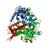
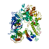
Journal: Plant Cell / Year: 2022
Title: Structure of plant RNA-DEPENDENT RNA POLYMERASE 2, an enzyme involved in small interfering RNA production.
Authors: Xuan Du / Zhenlin Yang / Alfredo Jose Florez Ariza / Qian Wang / Guohui Xie / Sisi Li / Jiamu Du /
Abstract: In plants, the biogenesis of small interfering RNA (siRNA) requires a family of RNA-dependent RNA polymerases that convert single-stranded RNA (ssRNA) into double-stranded RNA (dsRNA), which is ...In plants, the biogenesis of small interfering RNA (siRNA) requires a family of RNA-dependent RNA polymerases that convert single-stranded RNA (ssRNA) into double-stranded RNA (dsRNA), which is subsequently cleaved into defined lengths by Dicer endonucleases. Here, we determined the structure of maize (Zea mays) RNA-DEPENDENT RNA POLYMERASE 2 (ZmRDR2) in the closed and open conformations. The core catalytic region of ZmRDR2 possesses the canonical DNA-dependent RNA polymerase (DdRP) catalytic sites, pointing to a shared RNA production mechanism between DdRPs and plant RDR-family proteins. Apo-ZmRDR2 adopts a highly compact structure, representing an inactive closed conformation. By contrast, adding RNA induced a significant conformational change in the ZmRDR2 Head domain that opened the RNA binding tunnel, suggesting this is an active elongation conformation of ZmRDR2. Overall, our structural studies trapped both the active and inactive conformations of ZmRDR2, providing insights into the molecular mechanism of dsRNA synthesis during plant siRNA production.
History
DepositionDec 7, 2021Deposition site: PDBJ / Processing site: PDBJ
Revision 1.0Jun 8, 2022Provider: repository / Type: Initial release

-
Structure visualization

Structure viewerMolecule:
MolmilJmol/JSmol

Downloads & links

-
Assembly

Deposited unit
A: RNA-dependent RNA polymerase


Theoretical massNumber of molelcules
Total (without water)115,3921
Polymers115,3921
Non-polymers00
Water00
1


  • Idetical with deposited unit
  • defined by author
  • Evidence: gel filtration
TypeNameSymmetry operationNumber
identity operation1_555x,y,z1
Unit cell
Length a, b, c (Å)149.036, 149.036, 345.473
Angle α, β, γ (deg.)90.000, 90.000, 120.000
Int Tables number155
Space group name H-MH32

-
Components

#1: Protein RNA-dependent RNA polymerase / maize RDR2


Mass: 115392.172 Da / Num. of mol.: 1 / Mutation: E841A, K842A, E962A, E963A, E966A
Source method: isolated from a genetically manipulated source
Source: (gene. exp.) Zea mays (maize) / Gene: mop1, 100502460, ZEAMMB73_Zm00001d003378 / Plasmid: pFastBacHTB / Cell line (production host): Sf9 / Production host: 
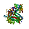
Spodoptera frugiperda (fall armyworm) / References: UniProt: Q19VG2, RNA-directed RNA polymerase

-
Experimental details

-
Experiment

ExperimentMethod: X-RAY DIFFRACTION / Number of used crystals: 1

-
Sample preparation

CrystalDensity Matthews: 3.23 Å3/Da / Density % sol: 61.87 %
Crystal growTemperature: 277 K / Method: vapor diffusion, sitting drop / pH: 6.5 / Details: 0.6M Sodium Citrate, 0.1M MES, pH 6.5

-
Data collection

DiffractionMean temperature: 100 K / Serial crystal experiment: N
Diffraction sourceSource: SYNCHROTRON / Site: SSRF / Beamline: BL19U1 / Wavelength: 0.9792 Å
DetectorType: DECTRIS PILATUS3 6M / Detector: PIXEL / Date: Oct 26, 2019
RadiationMonochromator: silicon crystal (111) / Protocol: SINGLE WAVELENGTH / Monochromatic (M) / Laue (L): M / Scattering type: x-ray
Radiation wavelengthWavelength: 0.9792 Å / Relative weight: 1
ReflectionResolution: 3.1→50 Å / Num. obs: 27101 / % possible obs: 99.7 % / Redundancy: 4.4 % / Biso Wilson estimate: 83.84 Å2 / Rmerge(I) obs: 0.124 / Rpim(I) all: 0.067 / Rrim(I) all: 0.141 / Χ2: 0.571 / Net I/σ(I): 3.7 / Num. measured all: 120266
Reflection shell

Diffraction-ID: 1

Resolution (Å)Redundancy (%)Rmerge(I) obsNum. unique obsCC1/2Rpim(I) allRrim(I) allΧ2% possible all
3.1-3.214.60.89426820.7490.4681.0110.4399.9
3.21-3.344.60.67526830.7930.3540.7640.43699.9
3.34-3.494.50.50126650.8840.2660.5690.47399.8
3.49-3.684.10.41226780.8670.2370.4770.97199.4
3.68-3.914.50.25727110.9420.1380.2930.706100
3.91-4.214.70.15526810.9860.080.1750.524100
4.21-4.634.60.127200.9920.0520.1130.59699.9
4.63-5.34.30.08427210.9920.0450.0960.59599.8
5.3-6.674.40.07627290.9920.0410.0860.5699.7
6.67-504.20.03628310.9980.020.0410.46898.7

-
Processing

Software
NameVersionClassification
PHENIX1.9_1692refinement
HKL-2000data scaling
PDB_EXTRACT3.27data extraction
HKL-3000data reduction
PHENIXphasing
RefinementMethod to determine structure: SAD / Resolution: 3.1→43.023 Å / SU ML: 0.46 / Cross valid method: THROUGHOUT / σ(F): 1.36 / Phase error: 28.74 / Stereochemistry target values: ML
RfactorNum. reflection% reflection
Rfree0.2703 1355 5.01 %
Rwork0.2301 25687 -
obs0.232 27042 99.5 %
Solvent computationShrinkage radii: 0.9 Å / VDW probe radii: 1.11 Å / Solvent model: FLAT BULK SOLVENT MODEL
Displacement parametersBiso max: 222.82 Å2 / Biso mean: 85.6438 Å2 / Biso min: 20 Å2
Refinement stepCycle: final / Resolution: 3.1→43.023 Å
ProteinNucleic acidLigandSolventTotal
Num. atoms7492 0 0 0 7492
Num. residues----944
Refine LS restraints
Refine-IDTypeDev idealNumber
X-RAY DIFFRACTIONf_bond_d0.0097658
X-RAY DIFFRACTIONf_angle_d1.29110352
X-RAY DIFFRACTIONf_chiral_restr0.0691138
X-RAY DIFFRACTIONf_plane_restr0.011332
X-RAY DIFFRACTIONf_dihedral_angle_d18.3452856
LS refinement shell

Refine-ID: X-RAY DIFFRACTION / Rfactor Rfree error: 0

Resolution (Å)Rfactor RfreeNum. reflection RfreeRfactor RworkNum. reflection Rwork% reflection obs (%)
3.1003-3.21110.36511220.3192558100
3.2111-3.33960.32271530.32182526100
3.3396-3.49150.34651370.29562522100
3.4915-3.67550.34751350.3277254099
3.6755-3.90570.30671320.28332577100
3.9057-4.2070.26061190.22592561100
4.207-4.62990.23321470.18742573100
4.6299-5.29880.25461520.19362568100
5.2988-6.6720.26531340.2252596100
6.672-430.22161240.1807266697
Refinement TLS params.

Method: refined / Refine-ID: X-RAY DIFFRACTION

IDL112)L122)L132)L222)L232)L332)S11 (Å °)S12 (Å °)S13 (Å °)S21 (Å °)S22 (Å °)S23 (Å °)S31 (Å °)S32 (Å °)S33 (Å °)T112)T122)T132)T222)T232)T332)Origin x (Å)Origin y (Å)Origin z (Å)
12.06550.1325-0.36081.427-0.15143.5568-0.04780.04380.3689-0.00480.0089-0.1489-0.65210.48990.01330.613-0.14850.00050.4547-0.0150.700274.7596186.4313360.495
20.6898-0.15890.05790.8917-0.00271.86930.0362-0.20010.00770.2511-0.02810.05830.201-0.05350.02680.60780.01620.0780.6041-0.03270.520454.6949160.9299379.4593
31.01180.73920.12282.96360.35672.13310.0332-0.4107-0.07390.3684-0.069-0.03260.50220.03770.00260.7670.06640.04630.5488-0.02740.454659.5376149.4957378.4385
42.1666-0.91750.04412.71690.45152.2585-0.06190.07550.46710.0161-0.06130.1503-0.1497-0.3380.10190.58260.01920.03490.56530.01190.688751.4601170.1454361.6183
Refinement TLS group
IDRefine-IDRefine TLS-IDSelection detailsAuth asym-IDAuth seq-ID
1X-RAY DIFFRACTION1chain 'A' and (resid 107 through 473 )A107 - 473
2X-RAY DIFFRACTION2chain 'A' and (resid 474 through 739 )A474 - 739
3X-RAY DIFFRACTION3chain 'A' and (resid 740 through 892 )A740 - 892
4X-RAY DIFFRACTION4chain 'A' and (resid 893 through 1111 )A893 - 1111

+
About Yorodumi

-
News

-
Feb 9, 2022. New format data for meta-information of EMDB entries

New format data for meta-information of EMDB entries

  • Version 3 of the EMDB header file is now the official format.
  • The previous official version 1.9 will be removed from the archive.

Related info.:EMDB header

External links:wwPDB to switch to version 3 of the EMDB data model

-
Aug 12, 2020. Covid-19 info

Covid-19 info

URL: https://pdbjlvh1.pdbj.org/emnavi/covid19.php

New page: Covid-19 featured information page in EM Navigator.

Related info.:Covid-19 info / Mar 5, 2020. Novel coronavirus structure data

+
Mar 5, 2020. Novel coronavirus structure data

Novel coronavirus structure data

Related info.:Yorodumi Speices / Aug 12, 2020. Covid-19 info

External links:COVID-19 featured content - PDBj / Molecule of the Month (242):Coronavirus Proteases

+
Jan 31, 2019. EMDB accession codes are about to change! (news from PDBe EMDB page)

EMDB accession codes are about to change! (news from PDBe EMDB page)

  • The allocation of 4 digits for EMDB accession codes will soon come to an end. Whilst these codes will remain in use, new EMDB accession codes will include an additional digit and will expand incrementally as the available range of codes is exhausted. The current 4-digit format prefixed with “EMD-” (i.e. EMD-XXXX) will advance to a 5-digit format (i.e. EMD-XXXXX), and so on. It is currently estimated that the 4-digit codes will be depleted around Spring 2019, at which point the 5-digit format will come into force.
  • The EM Navigator/Yorodumi systems omit the EMD- prefix.

Related info.:Q: What is EMD? / ID/Accession-code notation in Yorodumi/EM Navigator

External links:EMDB Accession Codes are Changing Soon! / Contact to PDBj

+
Jul 12, 2017. Major update of PDB

Major update of PDB

  • wwPDB released updated PDB data conforming to the new PDBx/mmCIF dictionary.
  • This is a major update changing the version number from 4 to 5, and with Remediation, in which all the entries are updated.
  • In this update, many items about electron microscopy experimental information are reorganized (e.g. em_software).
  • Now, EM Navigator and Yorodumi are based on the updated data.

External links:wwPDB Remediation / Enriched Model Files Conforming to OneDep Data Standards Now Available in the PDB FTP Archive

-
Yorodumi

Thousand views of thousand structures

  • Yorodumi is a browser for structure data from EMDB, PDB, SASBDB, etc.
  • This page is also the successor to EM Navigator detail page, and also detail information page/front-end page for Omokage search.
  • The word "yorodu" (or yorozu) is an old Japanese word meaning "ten thousand". "mi" (miru) is to see.

Related info.:EMDB / PDB / SASBDB / Comparison of 3 databanks / Yorodumi Search / Aug 31, 2016. New EM Navigator & Yorodumi / Yorodumi Papers / Jmol/JSmol / Function and homology information / Changes in new EM Navigator and Yorodumi

Read more