 Movie
Movie Controller

[English] 日本語
 Yorodumi
- PDB-7w65: Crystal structure of minor pilin TcpB from Vibrio cholerae comple... -
+ Open data

- Basic information
| Entry | Database: PDB / ID: 7w65 | |||||||||
|---|---|---|---|---|---|---|---|---|---|---|
| Title | Crystal structure of minor pilin TcpB from Vibrio cholerae complexed with secreted protein TcpF | |||||||||
 Components Components |
| |||||||||
 Keywords Keywords | CELL ADHESION / Type IVb pilus / Vibrio cholerae / Minor pilin. | |||||||||
| Function / homology | Vibrio cholerae toxin co-regulated pilus biosynthesis F / Vibrio cholerae toxin co-regulated pilus biosynthesis F, C-terminal / Vibrio cholerae toxin co-regulated pilus biosynthesis protein F / cell outer membrane / Toxin coregulated pilus biosynthesis protein F / Toxin-coregulated pilus biosynthesis protein B Function and homology information Function and homology information | |||||||||
| Biological species |   Vibrio cholerae (bacteria) Vibrio cholerae (bacteria) | |||||||||
| Method |  X-RAY DIFFRACTION / SYNCHROTRON / MOLECULAR REPLACEMENT / Resolution: 4.05 Å X-RAY DIFFRACTION / SYNCHROTRON / MOLECULAR REPLACEMENT / Resolution: 4.05 Å | |||||||||
 Authors Authors | Oki, H. / Kawahara, K. / Iimori, M. / Imoto, Y. / Maruno, T. / Uchiyama, S. / Muroga, Y. / Yoshida, A. / Yoshida, T. / Ohkubo, T. ...Oki, H. / Kawahara, K. / Iimori, M. / Imoto, Y. / Maruno, T. / Uchiyama, S. / Muroga, Y. / Yoshida, A. / Yoshida, T. / Ohkubo, T. / Matsuda, S. / Iida, T. / Nakamura, S. | |||||||||
| Funding support |  Japan, 2items Japan, 2items
| |||||||||
 Citation Citation | Journal: Sci Adv / Year: 2022 Title: Structural basis for the toxin-coregulated pilus-dependent secretion of Vibrio cholerae colonization factor. Authors: Oki, H. / Kawahara, K. / Iimori, M. / Imoto, Y. / Nishiumi, H. / Maruno, T. / Uchiyama, S. / Muroga, Y. / Yoshida, A. / Yoshida, T. / Ohkubo, T. / Matsuda, S. / Iida, T. / Nakamura, S. | |||||||||
| History |
|
- Structure visualization
| Structure viewer | Molecule: MolmilJmol/JSmol |
|---|
- Downloads & links
-Download
| PDBx/mmCIF format | 7w65.cif.gz | 414.1 KB | Display | PDBx/mmCIF format |
|---|---|---|---|---|
| PDB format | pdb7w65.ent.gz | 334.2 KB | Display | PDB format |
| PDBx/mmJSON format | 7w65.json.gz | Tree view | PDBx/mmJSON format | |
| Others |  Other downloads Other downloads |
-Validation report
| Arichive directory | https://data.pdbj.org/pub/pdb/validation_reports/w6/7w65ftp://data.pdbj.org/pub/pdb/validation_reports/w6/7w65 | HTTPS FTP |
|---|
-Related structure data
| Related structure data | 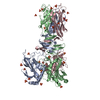 7w63SC  7w64C S: Starting model for refinement C: citing same article ( |
|---|---|
| Similar structure data |
-Links
 PDBj
PDBj- Assembly
| Deposited unit | 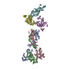
| ||||||||||||
|---|---|---|---|---|---|---|---|---|---|---|---|---|---|
| 1 |
| ||||||||||||
| Unit cell |
|
-Components
| #1: Protein | Mass: 43507.301 Da / Num. of mol.: 3 Source method: isolated from a genetically manipulated source Source: (gene. exp.) Vibrio cholerae (bacteria) / Gene: tcpB / Production host: #2: Protein | Mass: 35882.938 Da / Num. of mol.: 3 Source method: isolated from a genetically manipulated source Source: (gene. exp.) Vibrio cholerae (bacteria) / Gene: tcpF / Production host: Has protein modification | Y | |
|---|
-Experimental details
-Experiment
| Experiment | Method: X-RAY DIFFRACTION / Number of used crystals: 1 |
|---|
- Sample preparation
| Crystal | Density Matthews: 7.28 Å3/Da / Density % sol: 83.11 % Description: THE ENTRY CONTAINS FRIEDEL PAIRS IN I/F_PLUS/MINUS COLUMNS. |
|---|---|
| Crystal grow | Temperature: 273 K / Method: vapor diffusion, sitting drop Details: 0.4 M trisodium citrate, 4%(w/v) PEG3350, 10%(w/v) xylitol. |
-Data collection
| Diffraction | Mean temperature: 100 K / Serial crystal experiment: N | ||||||||||||||||||||||||||||||
|---|---|---|---|---|---|---|---|---|---|---|---|---|---|---|---|---|---|---|---|---|---|---|---|---|---|---|---|---|---|---|---|
| Diffraction source | Source: SYNCHROTRON / Site: SPring-8 / Beamline: BL38B1 / Wavelength: 1 Å | ||||||||||||||||||||||||||||||
| Detector | Type: DECTRIS PILATUS 6M / Detector: PIXEL / Date: Jan 30, 2019 | ||||||||||||||||||||||||||||||
| Radiation | Protocol: SINGLE WAVELENGTH / Monochromatic (M) / Laue (L): M / Scattering type: x-ray | ||||||||||||||||||||||||||||||
| Radiation wavelength | Wavelength: 1 Å / Relative weight: 1 | ||||||||||||||||||||||||||||||
| Reflection | Resolution: 4.05→49.21 Å / Num. obs: 57983 / % possible obs: 99.9 % / Redundancy: 19 % / CC1/2: 0.999 / Rmerge(I) obs: 0.17 / Rpim(I) all: 0.04 / Rrim(I) all: 0.175 / Net I/σ(I): 13.1 / Num. measured all: 1100587 / Scaling rejects: 33 | ||||||||||||||||||||||||||||||
| Reflection shell | Diffraction-ID: 1
|
- Processing
| Software |
| |||||||||||||||||||||||||||||||||||||||||||||||||||||||||||||||||||||||||||||||||||||||||||||||||||||||||||||||||||||||||||||||||||||||||||||||||||||||||||||||||||||||||||||||||||||||||||||||||||||||||||||||||||||||||
|---|---|---|---|---|---|---|---|---|---|---|---|---|---|---|---|---|---|---|---|---|---|---|---|---|---|---|---|---|---|---|---|---|---|---|---|---|---|---|---|---|---|---|---|---|---|---|---|---|---|---|---|---|---|---|---|---|---|---|---|---|---|---|---|---|---|---|---|---|---|---|---|---|---|---|---|---|---|---|---|---|---|---|---|---|---|---|---|---|---|---|---|---|---|---|---|---|---|---|---|---|---|---|---|---|---|---|---|---|---|---|---|---|---|---|---|---|---|---|---|---|---|---|---|---|---|---|---|---|---|---|---|---|---|---|---|---|---|---|---|---|---|---|---|---|---|---|---|---|---|---|---|---|---|---|---|---|---|---|---|---|---|---|---|---|---|---|---|---|---|---|---|---|---|---|---|---|---|---|---|---|---|---|---|---|---|---|---|---|---|---|---|---|---|---|---|---|---|---|---|---|---|---|---|---|---|---|---|---|---|---|---|---|---|---|---|---|---|---|
| Refinement | Method to determine structure: MOLECULAR REPLACEMENT Starting model: 7W63 Resolution: 4.05→31.83 Å / SU ML: 0.6315 / Cross valid method: FREE R-VALUE / σ(F): 1.33 / Phase error: 35.1153 Stereochemistry target values: GeoStd + Monomer Library + CDL v1.2
| |||||||||||||||||||||||||||||||||||||||||||||||||||||||||||||||||||||||||||||||||||||||||||||||||||||||||||||||||||||||||||||||||||||||||||||||||||||||||||||||||||||||||||||||||||||||||||||||||||||||||||||||||||||||||
| Solvent computation | Shrinkage radii: 0.9 Å / VDW probe radii: 1.11 Å / Solvent model: FLAT BULK SOLVENT MODEL | |||||||||||||||||||||||||||||||||||||||||||||||||||||||||||||||||||||||||||||||||||||||||||||||||||||||||||||||||||||||||||||||||||||||||||||||||||||||||||||||||||||||||||||||||||||||||||||||||||||||||||||||||||||||||
| Displacement parameters | Biso mean: 221.44 Å2 | |||||||||||||||||||||||||||||||||||||||||||||||||||||||||||||||||||||||||||||||||||||||||||||||||||||||||||||||||||||||||||||||||||||||||||||||||||||||||||||||||||||||||||||||||||||||||||||||||||||||||||||||||||||||||
| Refinement step | Cycle: LAST / Resolution: 4.05→31.83 Å
| |||||||||||||||||||||||||||||||||||||||||||||||||||||||||||||||||||||||||||||||||||||||||||||||||||||||||||||||||||||||||||||||||||||||||||||||||||||||||||||||||||||||||||||||||||||||||||||||||||||||||||||||||||||||||
| Refine LS restraints |
| |||||||||||||||||||||||||||||||||||||||||||||||||||||||||||||||||||||||||||||||||||||||||||||||||||||||||||||||||||||||||||||||||||||||||||||||||||||||||||||||||||||||||||||||||||||||||||||||||||||||||||||||||||||||||
| LS refinement shell |
|
