 Movie
Movie Controller

[English] 日本語
 Yorodumi
- PDB-7w5y: Cryo-EM structure of SoxS-dependent transcription activation comp... -
+ Open data

- Basic information
| Entry | Database: PDB / ID: 7w5y | ||||||
|---|---|---|---|---|---|---|---|
| Title | Cryo-EM structure of SoxS-dependent transcription activation complex with fpr promoter DNA | ||||||
 Components Components |
| ||||||
 Keywords Keywords | TRANSCRIPTION/DNA / bacterial RNA polymerase / TRANSCRIPTION-DNA COMPLEX | ||||||
| Function / homology |  Function and homology information Function and homology informationbacterial-type RNA polymerase holo enzyme binding / sigma factor antagonist complex / RNA polymerase complex / submerged biofilm formation / cellular response to cell envelope stress / regulation of DNA-templated transcription initiation / sigma factor activity / bacterial-type flagellum assembly / bacterial-type RNA polymerase core enzyme binding / cytosolic DNA-directed RNA polymerase complex ...bacterial-type RNA polymerase holo enzyme binding / sigma factor antagonist complex / RNA polymerase complex / submerged biofilm formation / cellular response to cell envelope stress / regulation of DNA-templated transcription initiation / sigma factor activity / bacterial-type flagellum assembly / bacterial-type RNA polymerase core enzyme binding / cytosolic DNA-directed RNA polymerase complex / bacterial-type flagellum-dependent cell motility / nitrate assimilation / cis-regulatory region sequence-specific DNA binding / regulation of DNA-templated transcription elongation / transcription elongation factor complex / DNA-directed RNA polymerase complex / transcription antitermination / cell motility / DNA-templated transcription initiation / ribonucleoside binding / DNA-directed RNA polymerase / DNA-directed RNA polymerase activity / response to heat / protein-containing complex assembly / sequence-specific DNA binding / intracellular iron ion homeostasis / protein dimerization activity / transcription cis-regulatory region binding / DNA-binding transcription factor activity / response to antibiotic / negative regulation of DNA-templated transcription / regulation of DNA-templated transcription / DNA-templated transcription / magnesium ion binding / DNA binding / zinc ion binding / membrane / cytoplasm / cytosol Similarity search - Function | ||||||
| Biological species |  | ||||||
| Method | ELECTRON MICROSCOPY / single particle reconstruction / cryo EM / Resolution: 4.2 Å | ||||||
 Authors Authors | Lin, W. / Feng, Y. | ||||||
| Funding support |  China, 1items China, 1items
| ||||||
 Citation Citation | Journal: Nucleic Acids Res / Year: 2022 Title: Structural basis of three different transcription activation strategies adopted by a single regulator SoxS. Authors: Jing Shi / Lu Wang / Aijia Wen / Fulin Wang / Yuqiong Zhang / Libing Yu / Fangfang Li / Yuanling Jin / Zhenzhen Feng / Jiacong Li / Yujiao Yang / Fei Gao / Yu Zhang / Yu Feng / Shuang Wang / ...Authors: Jing Shi / Lu Wang / Aijia Wen / Fulin Wang / Yuqiong Zhang / Libing Yu / Fangfang Li / Yuanling Jin / Zhenzhen Feng / Jiacong Li / Yujiao Yang / Fei Gao / Yu Zhang / Yu Feng / Shuang Wang / Wei Zhao / Wei Lin / Abstract: Transcription activation is established through extensive protein-protein and protein-DNA interactions that allow an activator to engage and remodel RNA polymerase. SoxS, a global transcription ...Transcription activation is established through extensive protein-protein and protein-DNA interactions that allow an activator to engage and remodel RNA polymerase. SoxS, a global transcription activator, diversely regulates subsets of stress response genes with different promoters, but the detailed SoxS-dependent transcription initiation mechanisms remain obscure. Here, we report cryo-EM structures of three SoxS-dependent transcription activation complexes (SoxS-TACI, SoxS-TACII and SoxS-TACIII) comprising of Escherichia coli RNA polymerase (RNAP), SoxS protein and three representative classes of SoxS-regulated promoters. The structures reveal that SoxS monomer orchestrates transcription initiation through specific interactions with the promoter DNA and different conserved domains of RNAP. In particular, SoxS is positioned in the opposite orientation in SoxS-TACIII to that in SoxS-TACI and SoxS-TACII, unveiling a novel mode of transcription activation. Strikingly, two universally conserved C-terminal domains of alpha subunit (αCTD) of RNAP associate with each other, bridging SoxS and region 4 of σ70. We show that SoxS interacts with RNAP directly and independently from DNA, remodeling the enzyme to activate transcription from cognate SoxS promoters while repressing transcription from UP-element containing promoters. Our data provide a comprehensive summary of SoxS-dependent promoter architectures and offer new insights into the αCTD contribution to transcription control in bacteria. | ||||||
| History |
|
- Structure visualization
| Structure viewer | Molecule: MolmilJmol/JSmol |
|---|
- Downloads & links
-Download
| PDBx/mmCIF format | 7w5y.cif.gz | 770.8 KB | Display | PDBx/mmCIF format |
|---|---|---|---|---|
| PDB format | pdb7w5y.ent.gz | 617.6 KB | Display | PDB format |
| PDBx/mmJSON format | 7w5y.json.gz | Tree view | PDBx/mmJSON format | |
| Others |  Other downloads Other downloads |
-Validation report
| Arichive directory | https://data.pdbj.org/pub/pdb/validation_reports/w5/7w5yftp://data.pdbj.org/pub/pdb/validation_reports/w5/7w5y | HTTPS FTP |
|---|
-Related structure data
| Related structure data | 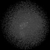 32324MC 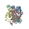 7w5wC 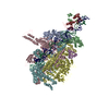 7w5xC M: map data used to model this data C: citing same article ( |
|---|---|
| Similar structure data |

-Links
 PDBj
PDBj


- Assembly
| Deposited unit | 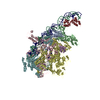
|
|---|---|
| 1 |
|
-Components
-DNA-directed RNA polymerase subunit ... , 4 types, 5 molecules ABCDE
| #1: Protein | Mass: 36558.680 Da / Num. of mol.: 2 Source method: isolated from a genetically manipulated source Source: (gene. exp.) #2: Protein | | Mass: 150804.922 Da / Num. of mol.: 1 / Mutation: D516V Source method: isolated from a genetically manipulated source Source: (gene. exp.) #3: Protein | | Mass: 155366.781 Da / Num. of mol.: 1 Source method: isolated from a genetically manipulated source Source: (gene. exp.) #4: Protein | | Mass: 10249.547 Da / Num. of mol.: 1 Source method: isolated from a genetically manipulated source Source: (gene. exp.) |
|---|
-Protein , 2 types, 2 molecules FK
| #5: Protein | Mass: 70352.242 Da / Num. of mol.: 1 Source method: isolated from a genetically manipulated source Source: (gene. exp.) |
|---|---|
| #8: Protein | Mass: 12931.844 Da / Num. of mol.: 1 Source method: isolated from a genetically manipulated source Source: (gene. exp.) |
-Fpr promoter DNA ... , 2 types, 2 molecules 12
| #6: DNA chain | Mass: 26637.020 Da / Num. of mol.: 1 / Source method: obtained synthetically / Source: (synth.) |
|---|---|
| #7: DNA chain | Mass: 26576.082 Da / Num. of mol.: 1 / Source method: obtained synthetically / Source: (synth.) |
-Experimental details
-Experiment
| Experiment | Method: ELECTRON MICROSCOPY |
|---|---|
| EM experiment | Aggregation state: PARTICLE / 3D reconstruction method: single particle reconstruction |
- Sample preparation
| Component |
| ||||||||||||||||||||||||
|---|---|---|---|---|---|---|---|---|---|---|---|---|---|---|---|---|---|---|---|---|---|---|---|---|---|
| Source (natural) |
| ||||||||||||||||||||||||
| Source (recombinant) | Organism: | ||||||||||||||||||||||||
| Buffer solution | pH: 7.5 | ||||||||||||||||||||||||
| Specimen | Embedding applied: NO / Shadowing applied: NO / Staining applied: NO / Vitrification applied: YES | ||||||||||||||||||||||||
| Vitrification | Cryogen name: ETHANE |
- Electron microscopy imaging
| Experimental equipment |  Model: Titan Krios / Image courtesy: FEI Company |
|---|---|
| Microscopy | Model: FEI TITAN KRIOS |
| Electron gun | Electron source:  FIELD EMISSION GUN / Accelerating voltage: 300 kV / Illumination mode: FLOOD BEAM FIELD EMISSION GUN / Accelerating voltage: 300 kV / Illumination mode: FLOOD BEAM |
| Electron lens | Mode: BRIGHT FIELD / Nominal defocus max: 2200 nm / Nominal defocus min: 1400 nm |
| Image recording | Electron dose: 52 e/Å2 / Detector mode: COUNTING / Film or detector model: GATAN K2 SUMMIT (4k x 4k) |
- Processing
| CTF correction | Type: NONE |
|---|---|
| 3D reconstruction | Resolution: 4.2 Å / Resolution method: FSC 0.143 CUT-OFF / Num. of particles: 74601 / Symmetry type: POINT |
