 Movie
Movie Controller


+ Open data

- Basic information
| Entry | Database: PDB / ID: 7w5r | ||||||
|---|---|---|---|---|---|---|---|
| Title | KRAS G12V and peptide complex | ||||||
 Components Components |
| ||||||
 Keywords Keywords | STRUCTURAL PROTEIN / Kras / hrev107 | ||||||
| Function / homology | small monomeric GTPase / Ca2+ pathway / P-loop containing nucleotide triphosphate hydrolases / Rossmann fold / 3-Layer(aba) Sandwich / Alpha Beta / GUANOSINE-5'-DIPHOSPHATE / Isoform 2B of GTPase KRas Function and homology information Function and homology information | ||||||
| Biological species |  Homo sapiens (human) Homo sapiens (human) | ||||||
| Method |  X-RAY DIFFRACTION / SYNCHROTRON / MOLECULAR REPLACEMENT / Resolution: 3.87 Å X-RAY DIFFRACTION / SYNCHROTRON / MOLECULAR REPLACEMENT / Resolution: 3.87 Å | ||||||
 Authors Authors | Kim, H.J. / Han, C.W. / Jang, S.B. | ||||||
| Funding support |  Korea, Republic Of, 1items Korea, Republic Of, 1items
| ||||||
 Citation Citation | Journal: Biochem.Biophys.Res.Commun. / Year: 2023 Title: Structural basis of the oncogenic KRAS mutant and GJ101 complex. Authors: Kim, H.J. / Han, C.W. / Jeong, M.S. / Jang, S.B. | ||||||
| History |
|
- Structure visualization
| Structure viewer | Molecule: MolmilJmol/JSmol |
|---|
- Downloads & links
-Download
| PDBx/mmCIF format | 7w5r.cif.gz | 470.3 KB | Display | PDBx/mmCIF format |
|---|---|---|---|---|
| PDB format | pdb7w5r.ent.gz | 319.7 KB | Display | PDB format |
| PDBx/mmJSON format | 7w5r.json.gz | Tree view | PDBx/mmJSON format | |
| Others |  Other downloads Other downloads |
-Validation report
| Arichive directory | https://data.pdbj.org/pub/pdb/validation_reports/w5/7w5rftp://data.pdbj.org/pub/pdb/validation_reports/w5/7w5r | HTTPS FTP |
|---|
-Related structure data
| Related structure data | 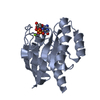 5uqwS S: Starting model for refinement |
|---|---|
| Similar structure data |
-Links
 PDBj
PDBj


- Assembly
| Deposited unit | 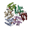
| ||||||||||||
|---|---|---|---|---|---|---|---|---|---|---|---|---|---|
| 1 |
| ||||||||||||
| Unit cell |
|
-Components
| #1: Protein | Mass: 20013.539 Da / Num. of mol.: 6 / Mutation: G12V Source method: isolated from a genetically manipulated source Source: (gene. exp.) Homo sapiens (human) / Gene: KRAS, KRAS2, RASK2 / Production host:  #2: Protein/peptide | Mass: 579.643 Da / Num. of mol.: 2 / Source method: obtained synthetically / Source: (synth.) Homo sapiens (human)#3: Chemical | ChemComp-MG /   Mass: 24.305 Da / Num. of mol.: 6 / Source method: obtained synthetically / Formula: Mg Mass: 24.305 Da / Num. of mol.: 6 / Source method: obtained synthetically / Formula: Mg#4: Chemical | ChemComp-GDP /   Type: RNA linking / Mass: 443.201 Da / Num. of mol.: 6 / Source method: obtained synthetically / Formula: C10H15N5O11P2 / Comment: GDP, energy-carrying molecule*YM Type: RNA linking / Mass: 443.201 Da / Num. of mol.: 6 / Source method: obtained synthetically / Formula: C10H15N5O11P2 / Comment: GDP, energy-carrying molecule*YMHas ligand of interest | N | Has protein modification | N | |
|---|
-Experimental details
-Experiment
| Experiment | Method: X-RAY DIFFRACTION / Number of used crystals: 1 |
|---|
- Sample preparation
| Crystal | Density Matthews: 2.11 Å3/Da / Density % sol: 41.58 % |
|---|---|
| Crystal grow | Temperature: 293.15 K / Method: vapor diffusion, hanging drop Details: polyethylene glycol 3350, 0.2 M potassium nitrate at pH 6.8 |
-Data collection
| Diffraction | Mean temperature: 100 K / Serial crystal experiment: N |
|---|---|
| Diffraction source | Source: SYNCHROTRON / Site: PAL/PLS / Beamline: 7A (6B, 6C1) / Wavelength: 0.979 Å |
| Detector | Type: ADSC QUANTUM 270 / Detector: CCD / Date: Jul 10, 2020 |
| Radiation | Protocol: SINGLE WAVELENGTH / Monochromatic (M) / Laue (L): M / Scattering type: x-ray |
| Radiation wavelength | Wavelength: 0.979 Å / Relative weight: 1 |
| Reflection | Resolution: 3.87→50 Å / Num. obs: 8581 / % possible obs: 99.64 % / Redundancy: 3.8 % / Biso Wilson estimate: 77.13 Å2 / Rmerge(I) obs: 0.088 / Net I/σ(I): 17.3 |
| Reflection shell | Resolution: 3.87→3.95 Å / Rmerge(I) obs: 0.1838 / Num. unique obs: 2674 |
- Processing
| Software |
| ||||||||||||||||||||||||||||||||||||||||
|---|---|---|---|---|---|---|---|---|---|---|---|---|---|---|---|---|---|---|---|---|---|---|---|---|---|---|---|---|---|---|---|---|---|---|---|---|---|---|---|---|---|
| Refinement | Method to determine structure: MOLECULAR REPLACEMENT Starting model: 5UQW Resolution: 3.87→35.76 Å / SU ML: 0.5682 / Cross valid method: FREE R-VALUE / σ(F): 1.53 / Phase error: 34.855
| ||||||||||||||||||||||||||||||||||||||||
| Solvent computation | Shrinkage radii: 0.9 Å / VDW probe radii: 1.11 Å / Solvent model: FLAT BULK SOLVENT MODEL | ||||||||||||||||||||||||||||||||||||||||
| Displacement parameters | Biso mean: 80.68 Å2 | ||||||||||||||||||||||||||||||||||||||||
| Refinement step | Cycle: LAST / Resolution: 3.87→35.76 Å
| ||||||||||||||||||||||||||||||||||||||||
| Refine LS restraints |
| ||||||||||||||||||||||||||||||||||||||||
| LS refinement shell | Resolution: 3.87→3.95 Å
| ||||||||||||||||||||||||||||||||||||||||
| Refinement TLS params. | Method: refined / Origin x: 9.71337819261 Å / Origin y: -19.2138395566 Å / Origin z: 25.4119434274 Å
| ||||||||||||||||||||||||||||||||||||||||
| Refinement TLS group | Selection details: all |
