 Movie
Movie Controller


+ Open data

- Basic information
| Entry | Database: PDB / ID: 7w3s | ||||||
|---|---|---|---|---|---|---|---|
| Title | The complex structure of Larg1-ADPr from Legionella pneumophila | ||||||
 Components Components | Type IV secretion protein Dot | ||||||
 Keywords Keywords | HYDROLASE / Legionella pneumophila effector complex / ADPr | ||||||
| Function / homology | Chem-AR6 / Type IV secretion protein Dot Function and homology information Function and homology information | ||||||
| Biological species |   Legionella pneumophila (bacteria) Legionella pneumophila (bacteria) | ||||||
| Method |  X-RAY DIFFRACTION / SYNCHROTRON / SAD / Resolution: 2.324 Å X-RAY DIFFRACTION / SYNCHROTRON / SAD / Resolution: 2.324 Å | ||||||
 Authors Authors | Ouyang, S. / Guan, H. / Li, P. | ||||||
| Funding support | 1items
| ||||||
 Citation Citation | Journal: mLife / Year: 2022 Title: Legionella pneumophila temporally regulates the activity of ADP/ATP translocases by reversible ADP-ribosylation. Authors: Fu, J. / Li, P. / Guan, H. / Huang, D. / Song, L. / Ouyang, S. / Luo, Z.Q. | ||||||
| History |
|
- Structure visualization
| Structure viewer | Molecule: MolmilJmol/JSmol |
|---|
- Downloads & links
-Download
| PDBx/mmCIF format | 7w3s.cif.gz | 353 KB | Display | PDBx/mmCIF format |
|---|---|---|---|---|
| PDB format | pdb7w3s.ent.gz | 286 KB | Display | PDB format |
| PDBx/mmJSON format | 7w3s.json.gz | Tree view | PDBx/mmJSON format | |
| Others |  Other downloads Other downloads |
-Validation report
| Summary document | 7w3s_validation.pdf.gz | 1.5 MB | Display | wwPDB validaton report |
|---|---|---|---|---|
| Full document | 7w3s_full_validation.pdf.gz | 1.5 MB | Display | |
| Data in XML | 7w3s_validation.xml.gz | 68.5 KB | Display | |
| Data in CIF | 7w3s_validation.cif.gz | 94.8 KB | Display | |
| Arichive directory | https://data.pdbj.org/pub/pdb/validation_reports/w3/7w3sftp://data.pdbj.org/pub/pdb/validation_reports/w3/7w3s | HTTPS FTP |
-Related structure data
| Similar structure data |
|---|
-Links
 PDBj
PDBj
- Assembly
| Deposited unit | 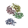
| ||||||||
|---|---|---|---|---|---|---|---|---|---|
| 1 | 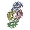
| ||||||||
| Unit cell |
|
-Components
| #1: Protein | Mass: 47037.523 Da / Num. of mol.: 4 Source method: isolated from a genetically manipulated source Source: (gene. exp.) Legionella pneumophila (bacteria) / Gene: C3927_15620 / Production host: #2: Chemical | ChemComp-AR6 / [(   Mass: 559.316 Da / Num. of mol.: 4 / Source method: obtained synthetically / Formula: C15H23N5O14P2 / Feature type: SUBJECT OF INVESTIGATION Mass: 559.316 Da / Num. of mol.: 4 / Source method: obtained synthetically / Formula: C15H23N5O14P2 / Feature type: SUBJECT OF INVESTIGATION#3: Water | ChemComp-HOH / |  Mass: 18.015 Da / Num. of mol.: 705 / Source method: isolated from a natural source / Formula: H2O Mass: 18.015 Da / Num. of mol.: 705 / Source method: isolated from a natural source / Formula: H2OHas ligand of interest | Y | Has protein modification | Y | |
|---|
-Experimental details
-Experiment
| Experiment | Method: X-RAY DIFFRACTION / Number of used crystals: 1 |
|---|
- Sample preparation
| Crystal | Density Matthews: 3 Å3/Da / Density % sol: 58.95 % |
|---|---|
| Crystal grow | Temperature: 289.15 K / Method: counter-diffusion Details: 0.2M Lithium sulfate, 0.1M Tris, pH 7.5, 5% polyethylene glycol 4,000 |
-Data collection
| Diffraction | Mean temperature: 100 K / Serial crystal experiment: N |
|---|---|
| Diffraction source | Source: SYNCHROTRON / Site: SSRF  / Beamline: BL17U1 / Wavelength: 0.97918 Å / Beamline: BL17U1 / Wavelength: 0.97918 Å |
| Detector | Type: DECTRIS EIGER X 16M / Detector: PIXEL / Date: Dec 18, 2020 |
| Radiation | Protocol: SINGLE WAVELENGTH / Monochromatic (M) / Laue (L): M / Scattering type: x-ray |
| Radiation wavelength | Wavelength: 0.97918 Å / Relative weight: 1 |
| Reflection | Resolution: 2.32→53.42 Å / Num. obs: 94202 / % possible obs: 97.54 % / Redundancy: 2 % / CC1/2: 0.998 / Net I/σ(I): 12.09 |
| Reflection shell | Resolution: 2.324→2.407 Å / Num. unique obs: 9251 / CC1/2: 0.773 |
- Processing
| Software |
| ||||||||||||||||||||||||||||||||||||||||||||||||||||||||||||||||||||||||||||||||||||||||||||||||||||||||||||||||||||||||||||||||||||||||||||||||||||||||||||||||||||||||||||||||||||||||||
|---|---|---|---|---|---|---|---|---|---|---|---|---|---|---|---|---|---|---|---|---|---|---|---|---|---|---|---|---|---|---|---|---|---|---|---|---|---|---|---|---|---|---|---|---|---|---|---|---|---|---|---|---|---|---|---|---|---|---|---|---|---|---|---|---|---|---|---|---|---|---|---|---|---|---|---|---|---|---|---|---|---|---|---|---|---|---|---|---|---|---|---|---|---|---|---|---|---|---|---|---|---|---|---|---|---|---|---|---|---|---|---|---|---|---|---|---|---|---|---|---|---|---|---|---|---|---|---|---|---|---|---|---|---|---|---|---|---|---|---|---|---|---|---|---|---|---|---|---|---|---|---|---|---|---|---|---|---|---|---|---|---|---|---|---|---|---|---|---|---|---|---|---|---|---|---|---|---|---|---|---|---|---|---|---|---|---|---|
| Refinement | Method to determine structure: SAD / Resolution: 2.324→53.416 Å / SU ML: 0.27 / Cross valid method: THROUGHOUT / σ(F): 1.34 / Phase error: 25.37 / Stereochemistry target values: ML
| ||||||||||||||||||||||||||||||||||||||||||||||||||||||||||||||||||||||||||||||||||||||||||||||||||||||||||||||||||||||||||||||||||||||||||||||||||||||||||||||||||||||||||||||||||||||||||
| Solvent computation | Shrinkage radii: 0.9 Å / VDW probe radii: 1.11 Å / Solvent model: FLAT BULK SOLVENT MODEL | ||||||||||||||||||||||||||||||||||||||||||||||||||||||||||||||||||||||||||||||||||||||||||||||||||||||||||||||||||||||||||||||||||||||||||||||||||||||||||||||||||||||||||||||||||||||||||
| Displacement parameters | Biso max: 89.79 Å2 / Biso mean: 42.2779 Å2 / Biso min: 18.52 Å2 | ||||||||||||||||||||||||||||||||||||||||||||||||||||||||||||||||||||||||||||||||||||||||||||||||||||||||||||||||||||||||||||||||||||||||||||||||||||||||||||||||||||||||||||||||||||||||||
| Refinement step | Cycle: final / Resolution: 2.324→53.416 Å
| ||||||||||||||||||||||||||||||||||||||||||||||||||||||||||||||||||||||||||||||||||||||||||||||||||||||||||||||||||||||||||||||||||||||||||||||||||||||||||||||||||||||||||||||||||||||||||
| Refine LS restraints |
| ||||||||||||||||||||||||||||||||||||||||||||||||||||||||||||||||||||||||||||||||||||||||||||||||||||||||||||||||||||||||||||||||||||||||||||||||||||||||||||||||||||||||||||||||||||||||||
| LS refinement shell | Refine-ID: X-RAY DIFFRACTION / Rfactor Rfree error: 0
|
