 Movie
Movie Controller


+ Open data

- Basic information
| Entry | Database: PDB / ID: 7w1x | ||||||
|---|---|---|---|---|---|---|---|
| Title | Crystal structure of AKR4C16 bound with NADPH | ||||||
 Components Components | AKR4-1 | ||||||
 Keywords Keywords | OXIDOREDUCTASE / Glyphosate resistance / GPJ degradation mechanism / aldo-keto reductase / structure-based engineering | ||||||
| Function / homology |  Function and homology information Function and homology information | ||||||
| Biological species |  Echinochloa colona (jungle rice) Echinochloa colona (jungle rice) | ||||||
| Method |  X-RAY DIFFRACTION / MOLECULAR REPLACEMENT / Resolution: 1.9 Å X-RAY DIFFRACTION / MOLECULAR REPLACEMENT / Resolution: 1.9 Å | ||||||
 Authors Authors | Li, H. / Yang, Y. / Hu, Y. / Chen, C.-C. / Huang, J.-W. / Min, J. / Dai, L. / Guo, R.-T. | ||||||
| Funding support |  China, 1items China, 1items
| ||||||
 Citation Citation | Journal: J Hazard Mater / Year: 2022 Title: Structural analysis and engineering of aldo-keto reductase from glyphosate-resistant Echinochloa colona Authors: Li, H. / Yang, Y. / Hu, Y. / Chen, C.C. / Huang, J.W. / Min, J. / Dai, L. / Guo, R.T. | ||||||
| History |
|
- Structure visualization
| Structure viewer | Molecule: MolmilJmol/JSmol |
|---|
- Downloads & links
-Download
| PDBx/mmCIF format | 7w1x.cif.gz | 147.6 KB | Display | PDBx/mmCIF format |
|---|---|---|---|---|
| PDB format | pdb7w1x.ent.gz | 113.1 KB | Display | PDB format |
| PDBx/mmJSON format | 7w1x.json.gz | Tree view | PDBx/mmJSON format | |
| Others |  Other downloads Other downloads |
-Validation report
| Arichive directory | https://data.pdbj.org/pub/pdb/validation_reports/w1/7w1xftp://data.pdbj.org/pub/pdb/validation_reports/w1/7w1x | HTTPS FTP |
|---|
-Related structure data
| Related structure data | 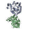 7f7jC 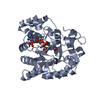 7f7kC  7f7lC 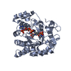 7f7mC 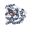 7w1wC  3h7uS S: Starting model for refinement C: citing same article ( |
|---|---|
| Similar structure data |
-Links
 PDBj
PDBj


- Assembly
| Deposited unit | 
| ||||||||
|---|---|---|---|---|---|---|---|---|---|
| 1 | 
| ||||||||
| 2 | 
| ||||||||
| Unit cell |
|
-Components
| #1: Protein | Mass: 35306.082 Da / Num. of mol.: 2 Source method: isolated from a genetically manipulated source Source: (gene. exp.) Echinochloa colona (jungle rice) / Production host:  #2: Chemical |   Mass: 745.421 Da / Num. of mol.: 2 / Source method: obtained synthetically / Formula: C21H30N7O17P3 / Feature type: SUBJECT OF INVESTIGATION Mass: 745.421 Da / Num. of mol.: 2 / Source method: obtained synthetically / Formula: C21H30N7O17P3 / Feature type: SUBJECT OF INVESTIGATION#3: Water | ChemComp-HOH / |  Mass: 18.015 Da / Num. of mol.: 520 / Source method: isolated from a natural source / Formula: H2O Mass: 18.015 Da / Num. of mol.: 520 / Source method: isolated from a natural source / Formula: H2OHas ligand of interest | Y | |
|---|
-Experimental details
-Experiment
| Experiment | Method: X-RAY DIFFRACTION / Number of used crystals: 1 |
|---|
- Sample preparation
| Crystal | Density Matthews: 1.96 Å3/Da / Density % sol: 37.18 % |
|---|---|
| Crystal grow | Temperature: 297 K / Method: evaporation / Details: 30 % PEG 4000, 0.2 M MgCl2, 0.1 M Tris pH 8.5, |
-Data collection
| Diffraction | Mean temperature: 100 K / Serial crystal experiment: N |
|---|---|
| Diffraction source | Source: LIQUID ANODE / Type: BRUKER METALJET / Wavelength: 1.34138 Å |
| Detector | Type: BRUKER PHOTON 100 / Detector: CMOS / Date: Oct 8, 2021 |
| Radiation | Protocol: SINGLE WAVELENGTH / Monochromatic (M) / Laue (L): M / Scattering type: x-ray |
| Radiation wavelength | Wavelength: 1.34138 Å / Relative weight: 1 |
| Reflection | Resolution: 1.9→34.88 Å / Num. obs: 42954 / % possible obs: 99.9 % / Redundancy: 6.96 % / CC1/2: 0.998 / Rmerge(I) obs: 0.0987 / Net I/σ(I): 11.58 |
| Reflection shell | Resolution: 1.9→1.93 Å / Rmerge(I) obs: 0.4476 / Mean I/σ(I) obs: 2.39 / Num. unique obs: 1965 |
- Processing
| Software |
| ||||||||||||||||||||||||||||||||||||||||||||||||||||||||||||
|---|---|---|---|---|---|---|---|---|---|---|---|---|---|---|---|---|---|---|---|---|---|---|---|---|---|---|---|---|---|---|---|---|---|---|---|---|---|---|---|---|---|---|---|---|---|---|---|---|---|---|---|---|---|---|---|---|---|---|---|---|---|
| Refinement | Method to determine structure: MOLECULAR REPLACEMENT Starting model: 3h7u Resolution: 1.9→34.88 Å / Cor.coef. Fo:Fc: 0.949 / Cor.coef. Fo:Fc free: 0.914 / SU B: 5.498 / SU ML: 0.155 / SU R Cruickshank DPI: 0.2095 / Cross valid method: THROUGHOUT / σ(F): 0 / ESU R: 0.21 / ESU R Free: 0.184 / Stereochemistry target values: MAXIMUM LIKELIHOOD Details: HYDROGENS HAVE BEEN ADDED IN THE RIDING POSITIONS U VALUES : REFINED INDIVIDUALLY
| ||||||||||||||||||||||||||||||||||||||||||||||||||||||||||||
| Solvent computation | Ion probe radii: 0.8 Å / Shrinkage radii: 0.8 Å / VDW probe radii: 1.2 Å / Solvent model: MASK | ||||||||||||||||||||||||||||||||||||||||||||||||||||||||||||
| Displacement parameters | Biso max: 76.6 Å2 / Biso mean: 24.845 Å2 / Biso min: 9.62 Å2
| ||||||||||||||||||||||||||||||||||||||||||||||||||||||||||||
| Refinement step | Cycle: final / Resolution: 1.9→34.88 Å
| ||||||||||||||||||||||||||||||||||||||||||||||||||||||||||||
| Refine LS restraints |
| ||||||||||||||||||||||||||||||||||||||||||||||||||||||||||||
| LS refinement shell | Resolution: 1.9→1.949 Å / Rfactor Rfree error: 0 / Total num. of bins used: 20
|
