 Movie
Movie Controller

[English] 日本語
 Yorodumi
- PDB-7w1c: Crystal structure of Klebsiella pneumoniae K1 capsule-specific po... -
+ Open data

- Basic information
| Entry | Database: PDB / ID: 7w1c | |||||||||
|---|---|---|---|---|---|---|---|---|---|---|
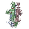
| Title | Crystal structure of Klebsiella pneumoniae K1 capsule-specific polysaccharide lyase in a P1 crystal form | |||||||||
 Components Components | K1 LYASE | |||||||||
 Keywords Keywords | VIRAL PROTEIN / BETA-HELIX / POLYSACCHARIDE LYASE / TAIL SPIKE PROTEIN | |||||||||
| Function / homology |  Function and homology information Function and homology informationsymbiont entry into host cell via disruption of host cell glycocalyx / Lyases / symbiont entry into host cell via disruption of host cell envelope / virus tail / adhesion receptor-mediated virion attachment to host cell / lyase activity Similarity search - Function | |||||||||
| Biological species |  Klebsiella phage NTUH-K2044-K1-1 (virus) Klebsiella phage NTUH-K2044-K1-1 (virus) | |||||||||
| Method |  X-RAY DIFFRACTION / SYNCHROTRON / MAD / Resolution: 1.48 Å X-RAY DIFFRACTION / SYNCHROTRON / MAD / Resolution: 1.48 Å | |||||||||
 Authors Authors | Tu, I.F. / Huang, K.F. / Wu, S.H. | |||||||||
| Funding support |  Taiwan, 2items Taiwan, 2items
| |||||||||
 Citation Citation | Journal: J.Biomed.Sci. / Year: 2022 Title: Structural and biological insights into Klebsiella pneumoniae surface polysaccharide degradation by a bacteriophage K1 lyase: implications for clinical use. Authors: Tu, I.F. / Lin, T.L. / Yang, F.L. / Lee, I.M. / Tu, W.L. / Liao, J.H. / Ko, T.P. / Wu, W.J. / Jan, J.T. / Ho, M.R. / Chou, C.Y. / Wang, A.H. / Wu, C.Y. / Wang, J.T. / Huang, K.F. / Wu, S.H. | |||||||||
| History |
|
- Structure visualization
| Structure viewer | Molecule: MolmilJmol/JSmol |
|---|
- Downloads & links
-Download
| PDBx/mmCIF format | 7w1c.cif.gz | 1.6 MB | Display | PDBx/mmCIF format |
|---|---|---|---|---|
| PDB format | pdb7w1c.ent.gz | Display | PDB format | |
| PDBx/mmJSON format | 7w1c.json.gz | Tree view | PDBx/mmJSON format | |
| Others |  Other downloads Other downloads |
-Validation report
| Arichive directory | https://data.pdbj.org/pub/pdb/validation_reports/w1/7w1cftp://data.pdbj.org/pub/pdb/validation_reports/w1/7w1c | HTTPS FTP |
|---|
-Related structure data

-Links
 PDBj
PDBj


- Assembly
| Deposited unit | 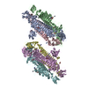
| ||||||||
|---|---|---|---|---|---|---|---|---|---|
| 1 | 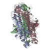
| ||||||||
| 2 | 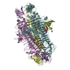
| ||||||||
| Unit cell |
|
-Components
| #1: Protein | Mass: 72130.633 Da / Num. of mol.: 6 / Mutation: D391A, D392A Source method: isolated from a genetically manipulated source Source: (gene. exp.) Klebsiella phage NTUH-K2044-K1-1 (virus)Plasmid: PET28A / Production host:  #2: Chemical | ChemComp-LMR / (   Mass: 134.087 Da / Num. of mol.: 7 / Source method: obtained synthetically / Formula: C4H6O5 / Feature type: SUBJECT OF INVESTIGATION Mass: 134.087 Da / Num. of mol.: 7 / Source method: obtained synthetically / Formula: C4H6O5 / Feature type: SUBJECT OF INVESTIGATION#3: Chemical | ChemComp-IMD /   Mass: 69.085 Da / Num. of mol.: 23 / Source method: obtained synthetically / Formula: C3H5N2 / Feature type: SUBJECT OF INVESTIGATION Mass: 69.085 Da / Num. of mol.: 23 / Source method: obtained synthetically / Formula: C3H5N2 / Feature type: SUBJECT OF INVESTIGATION#4: Chemical | ChemComp-GOL /   Mass: 92.094 Da / Num. of mol.: 18 / Source method: obtained synthetically / Formula: C3H8O3 / Feature type: SUBJECT OF INVESTIGATION Mass: 92.094 Da / Num. of mol.: 18 / Source method: obtained synthetically / Formula: C3H8O3 / Feature type: SUBJECT OF INVESTIGATION#5: Water | ChemComp-HOH / |  Mass: 18.015 Da / Num. of mol.: 4041 / Source method: isolated from a natural source / Formula: H2O Mass: 18.015 Da / Num. of mol.: 4041 / Source method: isolated from a natural source / Formula: H2OHas ligand of interest | Y | |
|---|
-Experimental details
-Experiment
| Experiment | Method: X-RAY DIFFRACTION / Number of used crystals: 1 |
|---|
- Sample preparation
| Crystal | Density Matthews: 2.45 Å3/Da / Density % sol: 49.8 % |
|---|---|
| Crystal grow | Temperature: 293 K / Method: vapor diffusion, sitting drop / pH: 6 Details: 15% (W/V) PEG 4000, 0.2 M IMIDAZOLE MALATE, PH 6.0, VAPOR DIFFUSION, SITTING DROP, TEMPERATURE 293K |
-Data collection
| Diffraction | Mean temperature: 100 K / Serial crystal experiment: N |
|---|---|
| Diffraction source | Source: SYNCHROTRON / Site: NSRRC / Beamline: BL15A1 / Wavelength: 1 Å |
| Detector | Type: RAYONIX MX300HE / Detector: CCD / Date: Oct 27, 2014 |
| Radiation | Monochromator: LN2-COOLED, FIXED-EXIT DOUBLE / Protocol: SINGLE WAVELENGTH / Monochromatic (M) / Laue (L): M / Scattering type: x-ray |
| Radiation wavelength | Wavelength: 1 Å / Relative weight: 1 |
| Reflection | Resolution: 1.48→30 Å / Num. obs: 656693 / % possible obs: 97 % / Observed criterion σ(I): 2 / Redundancy: 3.7 % / Rmerge(I) obs: 0.073 / Net I/σ(I): 25.44 |
| Reflection shell | Resolution: 1.48→1.53 Å / Redundancy: 3.7 % / Rmerge(I) obs: 0.741 / Mean I/σ(I) obs: 2.28 / Num. unique obs: 64500 / % possible all: 95.4 |
-Phasing
| Phasing | Method: MAD |
|---|
- Processing
| Software |
| |||||||||||||||||||||||||||||||||||||||||||||||||||||||||||||||||||||||||||||||||||||||||||||||||||||||||||||||||||||||||||||||||||||||||||||||||
|---|---|---|---|---|---|---|---|---|---|---|---|---|---|---|---|---|---|---|---|---|---|---|---|---|---|---|---|---|---|---|---|---|---|---|---|---|---|---|---|---|---|---|---|---|---|---|---|---|---|---|---|---|---|---|---|---|---|---|---|---|---|---|---|---|---|---|---|---|---|---|---|---|---|---|---|---|---|---|---|---|---|---|---|---|---|---|---|---|---|---|---|---|---|---|---|---|---|---|---|---|---|---|---|---|---|---|---|---|---|---|---|---|---|---|---|---|---|---|---|---|---|---|---|---|---|---|---|---|---|---|---|---|---|---|---|---|---|---|---|---|---|---|---|---|---|---|
| Refinement | Method to determine structure: MAD / Resolution: 1.48→30 Å / Cor.coef. Fo:Fc: 0.975 / Cor.coef. Fo:Fc free: 0.961 / SU B: 2.479 / SU ML: 0.042 / Cross valid method: THROUGHOUT / ESU R: 0.067 / ESU R Free: 0.065 / Stereochemistry target values: MAXIMUM LIKELIHOOD / Details: HYDROGENS HAVE BEEN ADDED IN THE RIDING POSITIONS
| |||||||||||||||||||||||||||||||||||||||||||||||||||||||||||||||||||||||||||||||||||||||||||||||||||||||||||||||||||||||||||||||||||||||||||||||||
| Solvent computation | Ion probe radii: 0.8 Å / Shrinkage radii: 0.8 Å / VDW probe radii: 1.2 Å / Solvent model: MASK | |||||||||||||||||||||||||||||||||||||||||||||||||||||||||||||||||||||||||||||||||||||||||||||||||||||||||||||||||||||||||||||||||||||||||||||||||
| Displacement parameters | Biso max: 125.19 Å2 / Biso mean: 21.99 Å2 / Biso min: 5.08 Å2
| |||||||||||||||||||||||||||||||||||||||||||||||||||||||||||||||||||||||||||||||||||||||||||||||||||||||||||||||||||||||||||||||||||||||||||||||||
| Refinement step | Cycle: final / Resolution: 1.48→30 Å
| |||||||||||||||||||||||||||||||||||||||||||||||||||||||||||||||||||||||||||||||||||||||||||||||||||||||||||||||||||||||||||||||||||||||||||||||||
| Refine LS restraints |
| |||||||||||||||||||||||||||||||||||||||||||||||||||||||||||||||||||||||||||||||||||||||||||||||||||||||||||||||||||||||||||||||||||||||||||||||||
| LS refinement shell | Resolution: 1.48→1.52 Å / Rfactor Rfree error: 0 / Total num. of bins used: 20
|
