 Movie
Movie Controller


+ Open data

- Basic information
| Entry | Database: PDB / ID: 7vx3 | |||||||||
|---|---|---|---|---|---|---|---|---|---|---|
| Title | OXA-58 crystal structure of acylated meropenem complex 2 | |||||||||
 Components Components | Beta-lactamase | |||||||||
 Keywords Keywords | HYDROLASE / OXA / multi-drug resistance / complex / degradation / ANTIBIOTIC | |||||||||
| Function / homology |  Function and homology information Function and homology informationpenicillin binding / antibiotic catabolic process / cell wall organization / beta-lactamase activity / beta-lactamase / response to antibiotic / plasma membrane Similarity search - Function | |||||||||
| Biological species |  Acinetobacter baumannii (bacteria) Acinetobacter baumannii (bacteria) | |||||||||
| Method |  X-RAY DIFFRACTION / MOLECULAR REPLACEMENT / Resolution: 1.8 Å X-RAY DIFFRACTION / MOLECULAR REPLACEMENT / Resolution: 1.8 Å | |||||||||
 Authors Authors | Saino, H. / Sugiyabu, T. / Miyano, M. | |||||||||
| Funding support |  Japan, 2items Japan, 2items
| |||||||||
 Citation Citation | Journal: To be published Title: OXA-58 crystal structure of acylated meropenem complex 2 Authors: Saino, H. / Sugiyabu, T. / Miyano, M. #1: Journal: PLoS One / Year: 2015Title: Crystal Structure of OXA-58 with the Substrate-Binding Cleft in a Closed State: Insights into the Mobility and Stability of the OXA-58 Structure. Authors: Saino, H. / Sugiyabu, T. / Ueno, G. / Yamamoto, M. / Ishii, Y. / Miyano, M. | |||||||||
| History |
|
- Structure visualization
| Structure viewer | Molecule: MolmilJmol/JSmol |
|---|
- Downloads & links
-Download
| PDBx/mmCIF format | 7vx3.cif.gz | 78 KB | Display | PDBx/mmCIF format |
|---|---|---|---|---|
| PDB format | pdb7vx3.ent.gz | 50.4 KB | Display | PDB format |
| PDBx/mmJSON format | 7vx3.json.gz | Tree view | PDBx/mmJSON format | |
| Others |  Other downloads Other downloads |
-Validation report
| Arichive directory | https://data.pdbj.org/pub/pdb/validation_reports/vx/7vx3ftp://data.pdbj.org/pub/pdb/validation_reports/vx/7vx3 | HTTPS FTP |
|---|
-Related structure data
| Related structure data |  7vx6C  7vviS S: Starting model for refinement C: citing same article ( |
|---|---|
| Similar structure data |
-Links
 PDBj
PDBj


- Assembly
| Deposited unit | 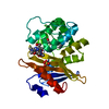
| ||||||||||||
|---|---|---|---|---|---|---|---|---|---|---|---|---|---|
| 1 |
| ||||||||||||
| Unit cell |
|
-Components
| #1: Protein | Mass: 31498.209 Da / Num. of mol.: 1 Source method: isolated from a genetically manipulated source Source: (gene. exp.) Acinetobacter baumannii (bacteria) / Gene: blaOXA-58, bla-oxa-58, bla-oxa58Production host: References: UniProt: Q2TR58, beta-lactamase |
|---|---|
| #2: Chemical | ChemComp-SO4 /   Mass: 96.063 Da / Num. of mol.: 1 / Source method: obtained synthetically / Formula: SO4 Mass: 96.063 Da / Num. of mol.: 1 / Source method: obtained synthetically / Formula: SO4 |
| #3: Chemical | ChemComp-MER / (  Mass: 385.478 Da / Num. of mol.: 1 / Source method: obtained synthetically / Formula: C17H27N3O5S / Feature type: SUBJECT OF INVESTIGATION / Comment: antibiotic*YM Mass: 385.478 Da / Num. of mol.: 1 / Source method: obtained synthetically / Formula: C17H27N3O5S / Feature type: SUBJECT OF INVESTIGATION / Comment: antibiotic*YM |
| #4: Chemical | ChemComp-DWZ / (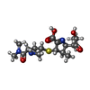  Mass: 385.478 Da / Num. of mol.: 1 / Source method: obtained synthetically / Formula: C17H27N3O5S / Feature type: SUBJECT OF INVESTIGATION / Comment: antibiotic*YM Mass: 385.478 Da / Num. of mol.: 1 / Source method: obtained synthetically / Formula: C17H27N3O5S / Feature type: SUBJECT OF INVESTIGATION / Comment: antibiotic*YM |
| #5: Water | ChemComp-HOH /  Mass: 18.015 Da / Num. of mol.: 206 / Source method: isolated from a natural source / Formula: H2O Mass: 18.015 Da / Num. of mol.: 206 / Source method: isolated from a natural source / Formula: H2O |
| Has ligand of interest | Y |
-Experimental details
-Experiment
| Experiment | Method: X-RAY DIFFRACTION / Number of used crystals: 1 |
|---|
- Sample preparation
| Crystal | Density Matthews: 2.07 Å3/Da / Density % sol: 40.55 % / Description: Thin plate |
|---|---|
| Crystal grow | Temperature: 298 K / Method: vapor diffusion, sitting drop / pH: 7.5 / Details: 0.1 M HEPES-Na (pH 7.5), 1.55 M LiSO4, NaCO3 |
-Data collection
| Diffraction | Mean temperature: 100 K / Ambient temp details: flash cooling / Serial crystal experiment: N |
|---|---|
| Diffraction source | Source: ROTATING ANODE / Type: RIGAKU MICROMAX-007 HF / Wavelength: 1.5418 Å |
| Detector | Type: RIGAKU RAXIS VII / Detector: IMAGE PLATE / Date: Jun 5, 2014 / Details: confocal mirror |
| Radiation | Monochromator: Ni-filter / Protocol: SINGLE WAVELENGTH / Monochromatic (M) / Laue (L): M / Scattering type: x-ray |
| Radiation wavelength | Wavelength: 1.5418 Å / Relative weight: 1 |
| Reflection | Resolution: 1.8→28.62 Å / Num. obs: 184659 / % possible obs: 99.95 % / Redundancy: 7.9 % / Biso Wilson estimate: 17.59 Å2 / CC1/2: 0.587 / CC star: 0.86 / Net I/σ(I): 65.48 |
| Reflection shell | Resolution: 1.8→1.864 Å / Redundancy: 7.6 % / Num. unique obs: 17814 / CC1/2: 0.371 / % possible all: 100 |
- Processing
| Software |
| |||||||||||||||||||||||||||||||||||||||||||||||||||||||||||||||||||||||||||||||||||||||||||||||||||||||||||||||||||||||||||||||||||||||||||||||||||
|---|---|---|---|---|---|---|---|---|---|---|---|---|---|---|---|---|---|---|---|---|---|---|---|---|---|---|---|---|---|---|---|---|---|---|---|---|---|---|---|---|---|---|---|---|---|---|---|---|---|---|---|---|---|---|---|---|---|---|---|---|---|---|---|---|---|---|---|---|---|---|---|---|---|---|---|---|---|---|---|---|---|---|---|---|---|---|---|---|---|---|---|---|---|---|---|---|---|---|---|---|---|---|---|---|---|---|---|---|---|---|---|---|---|---|---|---|---|---|---|---|---|---|---|---|---|---|---|---|---|---|---|---|---|---|---|---|---|---|---|---|---|---|---|---|---|---|---|---|
| Refinement | Method to determine structure: MOLECULAR REPLACEMENT Starting model: 7VVI Resolution: 1.8→28.62 Å / Cross valid method: FREE R-VALUE / σ(F): 1.13 / Phase error: 19.2616 Stereochemistry target values: GeoStd + Monomer Library + CDL v1.2
| |||||||||||||||||||||||||||||||||||||||||||||||||||||||||||||||||||||||||||||||||||||||||||||||||||||||||||||||||||||||||||||||||||||||||||||||||||
| Solvent computation | Shrinkage radii: 0.9 Å / VDW probe radii: 1.11 Å / Solvent model: FLAT BULK SOLVENT MODEL | |||||||||||||||||||||||||||||||||||||||||||||||||||||||||||||||||||||||||||||||||||||||||||||||||||||||||||||||||||||||||||||||||||||||||||||||||||
| Displacement parameters | Biso mean: 15.43 Å2 | |||||||||||||||||||||||||||||||||||||||||||||||||||||||||||||||||||||||||||||||||||||||||||||||||||||||||||||||||||||||||||||||||||||||||||||||||||
| Refinement step | Cycle: LAST / Resolution: 1.8→28.62 Å
| |||||||||||||||||||||||||||||||||||||||||||||||||||||||||||||||||||||||||||||||||||||||||||||||||||||||||||||||||||||||||||||||||||||||||||||||||||
| Refine LS restraints |
| |||||||||||||||||||||||||||||||||||||||||||||||||||||||||||||||||||||||||||||||||||||||||||||||||||||||||||||||||||||||||||||||||||||||||||||||||||
| LS refinement shell |
|
