 Movie
Movie Controller


+ Open data

- Basic information
| Entry | Database: PDB / ID: 7vvx | |||||||||||||||||||||
|---|---|---|---|---|---|---|---|---|---|---|---|---|---|---|---|---|---|---|---|---|---|---|
| Title | MmtN-SAH-Met complex | |||||||||||||||||||||
 Components Components | SAM-dependent methyltransferase | |||||||||||||||||||||
 Keywords Keywords | TRANSFERASE / SAM-dependent / Methyltransferase / complex / rossmann-like | |||||||||||||||||||||
| Function / homology | methyltransferase activity / methylation / S-adenosyl-L-methionine-dependent methyltransferase superfamily / METHIONINE / PHOSPHATE ION / S-ADENOSYL-L-HOMOCYSTEINE / SAM-dependent methyltransferase Function and homology information Function and homology information | |||||||||||||||||||||
| Biological species |  Roseovarius indicus (bacteria) Roseovarius indicus (bacteria) | |||||||||||||||||||||
| Method |  X-RAY DIFFRACTION / SYNCHROTRON / SAD / Resolution: 2.51 Å X-RAY DIFFRACTION / SYNCHROTRON / SAD / Resolution: 2.51 Å | |||||||||||||||||||||
 Authors Authors | Zhang, Y.Z. / Peng, M. / Li, C.Y. | |||||||||||||||||||||
| Funding support |  China, 6items China, 6items
| |||||||||||||||||||||
 Citation Citation | Journal: Nat Commun / Year: 2022 Title: Insights into methionine S-methylation in diverse organisms. Authors: Peng, M. / Li, C.Y. / Chen, X.L. / Williams, B.T. / Li, K. / Gao, Y.N. / Wang, P. / Wang, N. / Gao, C. / Zhang, S. / Schoelmerich, M.C. / Banfield, J.F. / Miller, J.B. / Le Brun, N.E. / Todd, J.D. / Zhang, Y.Z. | |||||||||||||||||||||
| History |
|
- Structure visualization
| Structure viewer | Molecule: MolmilJmol/JSmol |
|---|
- Downloads & links
-Download
| PDBx/mmCIF format | 7vvx.cif.gz | 167.9 KB | Display | PDBx/mmCIF format |
|---|---|---|---|---|
| PDB format | pdb7vvx.ent.gz | 131.8 KB | Display | PDB format |
| PDBx/mmJSON format | 7vvx.json.gz | Tree view | PDBx/mmJSON format | |
| Others |  Other downloads Other downloads |
-Validation report
| Arichive directory | https://data.pdbj.org/pub/pdb/validation_reports/vv/7vvxftp://data.pdbj.org/pub/pdb/validation_reports/vv/7vvx | HTTPS FTP |
|---|
-Related structure data

-Links
 PDBj
PDBj- Assembly
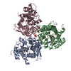
| Deposited unit | 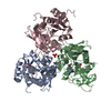
| ||||||||
|---|---|---|---|---|---|---|---|---|---|
| 1 |
| ||||||||
| Unit cell |
|
-Components
| #1: Protein | Mass: 32915.488 Da / Num. of mol.: 3 / Mutation: K141A/K143A/K146A Source method: isolated from a genetically manipulated source Source: (gene. exp.) Roseovarius indicus (bacteria) / Gene: XM52_07085 / Production host: #2: Chemical |   Type: L-peptide linking / Mass: 384.411 Da / Num. of mol.: 2 / Source method: obtained synthetically / Formula: C14H20N6O5S Type: L-peptide linking / Mass: 384.411 Da / Num. of mol.: 2 / Source method: obtained synthetically / Formula: C14H20N6O5S#3: Chemical | ChemComp-MET / |   Type: L-peptide linking / Mass: 149.211 Da / Num. of mol.: 1 / Source method: obtained synthetically / Formula: C5H11NO2S Type: L-peptide linking / Mass: 149.211 Da / Num. of mol.: 1 / Source method: obtained synthetically / Formula: C5H11NO2S#4: Chemical | ChemComp-PO4 / |   Mass: 94.971 Da / Num. of mol.: 1 / Source method: obtained synthetically / Formula: PO4 Mass: 94.971 Da / Num. of mol.: 1 / Source method: obtained synthetically / Formula: PO4#5: Water | ChemComp-HOH / |  Mass: 18.015 Da / Num. of mol.: 15 / Source method: isolated from a natural source / Formula: H2O Mass: 18.015 Da / Num. of mol.: 15 / Source method: isolated from a natural source / Formula: H2OHas ligand of interest | N | |
|---|
-Experimental details
-Experiment
| Experiment | Method: X-RAY DIFFRACTION / Number of used crystals: 1 |
|---|
- Sample preparation
| Crystal | Density Matthews: 2.99 Å3/Da / Density % sol: 63.18 % |
|---|---|
| Crystal grow | Temperature: 291.15 K / Method: vapor diffusion, hanging drop Details: 2% (vol/vol) PEG 400, 0.1M imidazole (pH 7.0), 24% (wt/vol) PEG MME 5000 |
-Data collection
| Diffraction | Mean temperature: 100 K / Serial crystal experiment: N |
|---|---|
| Diffraction source | Source: SYNCHROTRON / Site: SSRF / Beamline: BL17U1 / Wavelength: 0.97915 Å |
| Detector | Type: ADSC QUANTUM 315r / Detector: CCD / Date: Oct 21, 2019 |
| Radiation | Protocol: SINGLE WAVELENGTH / Monochromatic (M) / Laue (L): M / Scattering type: x-ray |
| Radiation wavelength | Wavelength: 0.97915 Å / Relative weight: 1 |
| Reflection | Resolution: 2.51→41.57 Å / Num. obs: 41239 / % possible obs: 99.7 % / Redundancy: 13 % / CC1/2: 0.999 / Rmerge(I) obs: 0.088 / Rpim(I) all: 0.025 / Rrim(I) all: 0.091 / Net I/σ(I): 17.2 |
| Reflection shell | Resolution: 2.51→2.6 Å / Redundancy: 13.6 % / Rmerge(I) obs: 1.755 / Mean I/σ(I) obs: 1.9 / Num. unique obs: 4072 / CC1/2: 0.81 / Rpim(I) all: 0.489 / Rrim(I) all: 1.823 / % possible all: 99.8 |
- Processing
| Software |
| ||||||||||||||||||||||||||||||||||||||||||||||||||||||||||||||||||||||||||||||||||||||||||||||||||||||||||||||||
|---|---|---|---|---|---|---|---|---|---|---|---|---|---|---|---|---|---|---|---|---|---|---|---|---|---|---|---|---|---|---|---|---|---|---|---|---|---|---|---|---|---|---|---|---|---|---|---|---|---|---|---|---|---|---|---|---|---|---|---|---|---|---|---|---|---|---|---|---|---|---|---|---|---|---|---|---|---|---|---|---|---|---|---|---|---|---|---|---|---|---|---|---|---|---|---|---|---|---|---|---|---|---|---|---|---|---|---|---|---|---|---|---|---|
| Refinement | Method to determine structure: SAD / Resolution: 2.51→41.57 Å / SU ML: 0.33 / Cross valid method: THROUGHOUT / σ(F): 1.34 / Phase error: 30.14 / Stereochemistry target values: ML
| ||||||||||||||||||||||||||||||||||||||||||||||||||||||||||||||||||||||||||||||||||||||||||||||||||||||||||||||||
| Solvent computation | Shrinkage radii: 0.9 Å / VDW probe radii: 1.11 Å / Solvent model: FLAT BULK SOLVENT MODEL | ||||||||||||||||||||||||||||||||||||||||||||||||||||||||||||||||||||||||||||||||||||||||||||||||||||||||||||||||
| Displacement parameters | Biso max: 144.85 Å2 / Biso mean: 76.4564 Å2 / Biso min: 20 Å2 | ||||||||||||||||||||||||||||||||||||||||||||||||||||||||||||||||||||||||||||||||||||||||||||||||||||||||||||||||
| Refinement step | Cycle: final / Resolution: 2.51→41.57 Å
| ||||||||||||||||||||||||||||||||||||||||||||||||||||||||||||||||||||||||||||||||||||||||||||||||||||||||||||||||
| LS refinement shell | Refine-ID: X-RAY DIFFRACTION / Rfactor Rfree error: 0 / Total num. of bins used: 15
|
