 Movie
Movie Controller

[English] 日本語
 Yorodumi
- PDB-7vua: Anaerobic hydroxyproline degradation involving C-N cleavage by a ... -
+ Open data

- Basic information
| Entry | Database: PDB / ID: 7vua | ||||||
|---|---|---|---|---|---|---|---|
| Title | Anaerobic hydroxyproline degradation involving C-N cleavage by a glycyl radical enzyme | ||||||
 Components Components | HplG | ||||||
 Keywords Keywords | LYASE / C-N-lyase | ||||||
| Function / homology | Pyruvate formate lyase domain / Pyruvate formate lyase-like / Pyruvate formate-lyase domain profile. / Glycine radical / Glycine radical domain / Glycine radical domain profile. / catalytic activity / (4S)-4-hydroxy-D-proline / Uncharacterized protein Function and homology information Function and homology information | ||||||
| Biological species |  Clostridiales bacterium (bacteria) Clostridiales bacterium (bacteria) | ||||||
| Method |  X-RAY DIFFRACTION / SYNCHROTRON / MOLECULAR REPLACEMENT / Resolution: 2.695 Å X-RAY DIFFRACTION / SYNCHROTRON / MOLECULAR REPLACEMENT / Resolution: 2.695 Å | ||||||
 Authors Authors | Duan, Y. / Lu, Q. / Yuchi, Z. / Zhang, Y. | ||||||
| Funding support |  China, 1items China, 1items
| ||||||
 Citation Citation | Journal: J.Am.Chem.Soc. / Year: 2022 Title: Anaerobic Hydroxyproline Degradation Involving C-N Cleavage by a Glycyl Radical Enzyme. Authors: Duan, Y. / Wei, Y. / Xing, M. / Liu, J. / Jiang, L. / Lu, Q. / Liu, X. / Liu, Y. / Ang, E.L. / Liao, R.Z. / Yuchi, Z. / Zhao, H. / Zhang, Y. | ||||||
| History |
|
- Structure visualization
| Structure viewer | Molecule: MolmilJmol/JSmol |
|---|
- Downloads & links
-Download
| PDBx/mmCIF format | 7vua.cif.gz | 322.1 KB | Display | PDBx/mmCIF format |
|---|---|---|---|---|
| PDB format | pdb7vua.ent.gz | 256.7 KB | Display | PDB format |
| PDBx/mmJSON format | 7vua.json.gz | Tree view | PDBx/mmJSON format | |
| Others |  Other downloads Other downloads |
-Validation report
| Arichive directory | https://data.pdbj.org/pub/pdb/validation_reports/vu/7vuaftp://data.pdbj.org/pub/pdb/validation_reports/vu/7vua | HTTPS FTP |
|---|
-Related structure data
| Similar structure data |
|---|
-Links
 PDBj
PDBj- Assembly
| Deposited unit | 
| ||||||||
|---|---|---|---|---|---|---|---|---|---|
| 1 |
| ||||||||
| Unit cell |
|
-Components
| #1: Protein | Mass: 88971.680 Da / Num. of mol.: 2 / Mutation: E106A/E107A/E108A Source method: isolated from a genetically manipulated source Source: (gene. exp.) Clostridiales bacterium (bacteria) / Gene: DBY07_03870 / Production host: #2: Chemical | 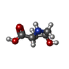  Type: D-peptide linking / Mass: 131.130 Da / Num. of mol.: 2 / Source method: obtained synthetically / Formula: C5H9NO3 / Feature type: SUBJECT OF INVESTIGATION Type: D-peptide linking / Mass: 131.130 Da / Num. of mol.: 2 / Source method: obtained synthetically / Formula: C5H9NO3 / Feature type: SUBJECT OF INVESTIGATION#3: Water | ChemComp-HOH / |  Mass: 18.015 Da / Num. of mol.: 352 / Source method: isolated from a natural source / Formula: H2O Mass: 18.015 Da / Num. of mol.: 352 / Source method: isolated from a natural source / Formula: H2OHas ligand of interest | Y | |
|---|
-Experimental details
-Experiment
| Experiment | Method: X-RAY DIFFRACTION / Number of used crystals: 1 |
|---|
- Sample preparation
| Crystal | Density Matthews: 3.03 Å3/Da / Density % sol: 59.42 % |
|---|---|
| Crystal grow | Temperature: 291.15 K / Method: vapor diffusion, sitting drop Details: 0.1M HEPES pH 7.5, 10% (w/v) PEG 8000, 8% (v/v) Ethylene glycol |
-Data collection
| Diffraction | Mean temperature: 100 K / Serial crystal experiment: N | ||||||||||||||||||||||||||||||||||||||||||||||||||||||||||||||||||||||||||||||||||||||||||||||||||||||||||||||||||||||||||||||||||||||||||||||||||||||||||||||||||||||||
|---|---|---|---|---|---|---|---|---|---|---|---|---|---|---|---|---|---|---|---|---|---|---|---|---|---|---|---|---|---|---|---|---|---|---|---|---|---|---|---|---|---|---|---|---|---|---|---|---|---|---|---|---|---|---|---|---|---|---|---|---|---|---|---|---|---|---|---|---|---|---|---|---|---|---|---|---|---|---|---|---|---|---|---|---|---|---|---|---|---|---|---|---|---|---|---|---|---|---|---|---|---|---|---|---|---|---|---|---|---|---|---|---|---|---|---|---|---|---|---|---|---|---|---|---|---|---|---|---|---|---|---|---|---|---|---|---|---|---|---|---|---|---|---|---|---|---|---|---|---|---|---|---|---|---|---|---|---|---|---|---|---|---|---|---|---|---|---|---|---|
| Diffraction source | Source: SYNCHROTRON / Site: SSRF / Beamline: BL18U1 / Wavelength: 0.9795 Å | ||||||||||||||||||||||||||||||||||||||||||||||||||||||||||||||||||||||||||||||||||||||||||||||||||||||||||||||||||||||||||||||||||||||||||||||||||||||||||||||||||||||||
| Detector | Type: DECTRIS PILATUS3 6M / Detector: PIXEL / Date: Jan 25, 2021 | ||||||||||||||||||||||||||||||||||||||||||||||||||||||||||||||||||||||||||||||||||||||||||||||||||||||||||||||||||||||||||||||||||||||||||||||||||||||||||||||||||||||||
| Radiation | Protocol: SINGLE WAVELENGTH / Monochromatic (M) / Laue (L): M / Scattering type: x-ray | ||||||||||||||||||||||||||||||||||||||||||||||||||||||||||||||||||||||||||||||||||||||||||||||||||||||||||||||||||||||||||||||||||||||||||||||||||||||||||||||||||||||||
| Radiation wavelength | Wavelength: 0.9795 Å / Relative weight: 1 | ||||||||||||||||||||||||||||||||||||||||||||||||||||||||||||||||||||||||||||||||||||||||||||||||||||||||||||||||||||||||||||||||||||||||||||||||||||||||||||||||||||||||
| Reflection | Resolution: 2.695→50 Å / Num. obs: 59889 / % possible obs: 100 % / Redundancy: 13.3 % / Rmerge(I) obs: 0.141 / Rpim(I) all: 0.04 / Rrim(I) all: 0.147 / Χ2: 0.677 / Net I/σ(I): 3.6 | ||||||||||||||||||||||||||||||||||||||||||||||||||||||||||||||||||||||||||||||||||||||||||||||||||||||||||||||||||||||||||||||||||||||||||||||||||||||||||||||||||||||||
| Reflection shell | Diffraction-ID: 1 / % possible all: 100
|
- Processing
| Software |
| ||||||||||||||||||||||||||||||||||||||||||||||||||||||||||||||||||||||||||||||||||||||||||
|---|---|---|---|---|---|---|---|---|---|---|---|---|---|---|---|---|---|---|---|---|---|---|---|---|---|---|---|---|---|---|---|---|---|---|---|---|---|---|---|---|---|---|---|---|---|---|---|---|---|---|---|---|---|---|---|---|---|---|---|---|---|---|---|---|---|---|---|---|---|---|---|---|---|---|---|---|---|---|---|---|---|---|---|---|---|---|---|---|---|---|---|
| Refinement | Method to determine structure: MOLECULAR REPLACEMENT Starting model: Phyre2 prediction struction Resolution: 2.695→32.142 Å / SU ML: 0.35 / Cross valid method: THROUGHOUT / σ(F): 0 / Phase error: 25.03 / Stereochemistry target values: ML
| ||||||||||||||||||||||||||||||||||||||||||||||||||||||||||||||||||||||||||||||||||||||||||
| Solvent computation | Shrinkage radii: 0.9 Å / VDW probe radii: 1.11 Å / Solvent model: FLAT BULK SOLVENT MODEL | ||||||||||||||||||||||||||||||||||||||||||||||||||||||||||||||||||||||||||||||||||||||||||
| Displacement parameters | Biso max: 114.66 Å2 / Biso mean: 46.5257 Å2 / Biso min: 16.16 Å2 | ||||||||||||||||||||||||||||||||||||||||||||||||||||||||||||||||||||||||||||||||||||||||||
| Refinement step | Cycle: final / Resolution: 2.695→32.142 Å
| ||||||||||||||||||||||||||||||||||||||||||||||||||||||||||||||||||||||||||||||||||||||||||
| LS refinement shell | Refine-ID: X-RAY DIFFRACTION / Rfactor Rfree error: 0
|
