 Movie
Movie Controller


+ Open data

- Basic information
| Entry | Database: PDB / ID: 7vtw | |||||||||
|---|---|---|---|---|---|---|---|---|---|---|
| Title | Crystal structure of PDE8A catalytic domain in complex with 17 | |||||||||
 Components Components | High affinity cAMP-specific and IBMX-insensitive 3',5'-cyclic phosphodiesterase 8A | |||||||||
 Keywords Keywords | HYDROLASE / cAMP-specific / Selective PDE8A inhibitor | |||||||||
| Function / homology |  Function and homology information Function and homology information3',5'-cyclic-AMP phosphodiesterase / : / cAMP catabolic process / 3',5'-cyclic-GMP phosphodiesterase activity / 3',5'-cyclic-AMP phosphodiesterase activity / protein kinase activator activity / cellular response to epidermal growth factor stimulus / kinase binding / G alpha (s) signalling events / positive regulation of ERK1 and ERK2 cascade ...3',5'-cyclic-AMP phosphodiesterase / : / cAMP catabolic process / 3',5'-cyclic-GMP phosphodiesterase activity / 3',5'-cyclic-AMP phosphodiesterase activity / protein kinase activator activity / cellular response to epidermal growth factor stimulus / kinase binding / G alpha (s) signalling events / positive regulation of ERK1 and ERK2 cascade / regulation of DNA-templated transcription / extracellular exosome / metal ion binding / cytosol Similarity search - Function | |||||||||
| Biological species |  Homo sapiens (human) Homo sapiens (human) | |||||||||
| Method |  X-RAY DIFFRACTION / MOLECULAR REPLACEMENT / Resolution: 2.79971328057 Å X-RAY DIFFRACTION / MOLECULAR REPLACEMENT / Resolution: 2.79971328057 Å | |||||||||
 Authors Authors | Wu, X.-N. / Zhou, Q. / Huang, Y.-D. / Li, Z. / Wu, Y. / Luo, H.-B. | |||||||||
| Funding support |  China, 2items China, 2items
| |||||||||
 Citation Citation | Journal: To Be Published Title: Structure-Based Discovery of Orally Efficient PDE8 Inhibitors for the Treatment of Vascular Dementia Authors: Wu, X.-N. / Zhou, Q. / Huang, Y.-D. / Li, Z. / Wu, Y. / Luo, H.-B. | |||||||||
| History |
|
- Structure visualization
| Structure viewer | Molecule: MolmilJmol/JSmol |
|---|
- Downloads & links
-Download
| PDBx/mmCIF format | 7vtw.cif.gz | 85.1 KB | Display | PDBx/mmCIF format |
|---|---|---|---|---|
| PDB format | pdb7vtw.ent.gz | 60 KB | Display | PDB format |
| PDBx/mmJSON format | 7vtw.json.gz | Tree view | PDBx/mmJSON format | |
| Others |  Other downloads Other downloads |
-Validation report
| Arichive directory | https://data.pdbj.org/pub/pdb/validation_reports/vt/7vtwftp://data.pdbj.org/pub/pdb/validation_reports/vt/7vtw | HTTPS FTP |
|---|
-Related structure data
| Related structure data |  7vslC  7vtvC  7vtxC  3ecmS S: Starting model for refinement C: citing same article ( |
|---|---|
| Similar structure data |
-Links
 PDBj
PDBj


- Assembly
| Deposited unit | 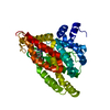
| ||||||||||||
|---|---|---|---|---|---|---|---|---|---|---|---|---|---|
| 1 |
| ||||||||||||
| Unit cell |
|
-Components
| #1: Protein | Mass: 39090.020 Da / Num. of mol.: 1 Source method: isolated from a genetically manipulated source Source: (gene. exp.) Homo sapiens (human) / Gene: PDE8A / Production host:  References: UniProt: O60658, 3',5'-cyclic-AMP phosphodiesterase |
|---|---|
| #2: Chemical | ChemComp-ZN /   Mass: 65.409 Da / Num. of mol.: 1 / Source method: obtained synthetically / Formula: Zn / Feature type: SUBJECT OF INVESTIGATION Mass: 65.409 Da / Num. of mol.: 1 / Source method: obtained synthetically / Formula: Zn / Feature type: SUBJECT OF INVESTIGATION |
| #3: Chemical | ChemComp-MG /   Mass: 24.305 Da / Num. of mol.: 1 / Source method: obtained synthetically / Formula: Mg / Feature type: SUBJECT OF INVESTIGATION Mass: 24.305 Da / Num. of mol.: 1 / Source method: obtained synthetically / Formula: Mg / Feature type: SUBJECT OF INVESTIGATION |
| #4: Chemical | ChemComp-807 / 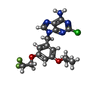  Mass: 397.807 Da / Num. of mol.: 1 / Source method: obtained synthetically / Formula: C17H18ClF2N5O2 / Feature type: SUBJECT OF INVESTIGATION Mass: 397.807 Da / Num. of mol.: 1 / Source method: obtained synthetically / Formula: C17H18ClF2N5O2 / Feature type: SUBJECT OF INVESTIGATION |
| #5: Water | ChemComp-HOH /  Mass: 18.015 Da / Num. of mol.: 37 / Source method: isolated from a natural source / Formula: H2O Mass: 18.015 Da / Num. of mol.: 37 / Source method: isolated from a natural source / Formula: H2O |
| Has ligand of interest | Y |
| Has protein modification | Y |
-Experimental details
-Experiment
| Experiment | Method: X-RAY DIFFRACTION / Number of used crystals: 1 |
|---|
- Sample preparation
| Crystal | Density Matthews: 3.24 Å3/Da / Density % sol: 62.03 % |
|---|---|
| Crystal grow | Temperature: 277 K / Method: vapor diffusion, hanging drop Details: 100mM Cacodylate Sodium pH 6.5, 15% Isopropanol, 30% Ethylene Glycol, 11% PEG 3350 |
-Data collection
| Diffraction | Mean temperature: 100 K / Serial crystal experiment: N |
|---|---|
| Diffraction source | Source: SEALED TUBE / Type: OXFORD DIFFRACTION NOVA / Wavelength: 1.5406 Å |
| Detector | Type: OXFORD ONYX CCD / Detector: CCD / Date: Jul 26, 2020 |
| Radiation | Protocol: SINGLE WAVELENGTH / Monochromatic (M) / Laue (L): M / Scattering type: x-ray |
| Radiation wavelength | Wavelength: 1.5406 Å / Relative weight: 1 |
| Reflection | Resolution: 2.799→24.173 Å / Num. obs: 12812 / % possible obs: 99.5 % / Redundancy: 5.2 % / Biso Wilson estimate: 45.9311676557 Å2 / Rmerge(I) obs: 0.103 / Net I/σ(I): 18.73 |
| Reflection shell | Resolution: 2.8→2.9 Å / Rmerge(I) obs: 0.276 / Num. unique obs: 1283 |
- Processing
| Software |
| ||||||||||||||||||||||||||||||||||||||||||
|---|---|---|---|---|---|---|---|---|---|---|---|---|---|---|---|---|---|---|---|---|---|---|---|---|---|---|---|---|---|---|---|---|---|---|---|---|---|---|---|---|---|---|---|
| Refinement | Method to determine structure: MOLECULAR REPLACEMENT Starting model: 3ECM Resolution: 2.79971328057→24.1726451871 Å / SU ML: 0.420550145062 / Cross valid method: THROUGHOUT / σ(F): 1.96981342147 / Phase error: 29.0159321752 Stereochemistry target values: GeoStd + Monomer Library + CDL v1.2
| ||||||||||||||||||||||||||||||||||||||||||
| Solvent computation | Shrinkage radii: 0.9 Å / VDW probe radii: 1.11 Å / Solvent model: FLAT BULK SOLVENT MODEL | ||||||||||||||||||||||||||||||||||||||||||
| Displacement parameters | Biso mean: 46.2347383513 Å2 | ||||||||||||||||||||||||||||||||||||||||||
| Refinement step | Cycle: LAST / Resolution: 2.79971328057→24.1726451871 Å
| ||||||||||||||||||||||||||||||||||||||||||
| Refine LS restraints |
| ||||||||||||||||||||||||||||||||||||||||||
| LS refinement shell |
|
