 Movie
Movie Controller

[English] 日本語
 Yorodumi
- PDB-7vs7: Crystal structure of the ectodomain of OsCERK1 in complex with ch... -
+ Open data

- Basic information
| Entry | Database: PDB / ID: 7vs7 | ||||||
|---|---|---|---|---|---|---|---|
| Title | Crystal structure of the ectodomain of OsCERK1 in complex with chitin hexamer | ||||||
 Components Components | Chitin elicitor receptor kinase 1 | ||||||
 Keywords Keywords | TRANSFERASE / CERK1 / chitin / PAMP / immunity / symbiosis / immune receptor / ANTIFUNGAL PROTEIN | ||||||
| Function / homology |  Function and homology information Function and homology informationtransmembrane receptor protein kinase activity / chitin binding / non-specific serine/threonine protein kinase / innate immune response / protein serine kinase activity / protein serine/threonine kinase activity / ATP binding / plasma membrane Similarity search - Function | ||||||
| Biological species |  | ||||||
| Method |  X-RAY DIFFRACTION / SYNCHROTRON / MOLECULAR REPLACEMENT / Resolution: 2.015 Å X-RAY DIFFRACTION / SYNCHROTRON / MOLECULAR REPLACEMENT / Resolution: 2.015 Å | ||||||
 Authors Authors | Li, X. | ||||||
| Funding support | 1items
| ||||||
 Citation Citation | Journal: J Integr Plant Biol / Year: 2023 Title: Structural insight into chitin perception by chitin elicitor receptor kinase 1 of Oryza sativa. Authors: Xu, L. / Wang, J. / Xiao, Y. / Han, Z. / Chai, J. | ||||||
| History |
|
- Structure visualization
| Structure viewer | Molecule: MolmilJmol/JSmol |
|---|
- Downloads & links
-Download
| PDBx/mmCIF format | 7vs7.cif.gz | 98.4 KB | Display | PDBx/mmCIF format |
|---|---|---|---|---|
| PDB format | pdb7vs7.ent.gz | 73.6 KB | Display | PDB format |
| PDBx/mmJSON format | 7vs7.json.gz | Tree view | PDBx/mmJSON format | |
| Others |  Other downloads Other downloads |
-Validation report
| Summary document | 7vs7_validation.pdf.gz | 1.6 MB | Display | wwPDB validaton report |
|---|---|---|---|---|
| Full document | 7vs7_full_validation.pdf.gz | 1.6 MB | Display | |
| Data in XML | 7vs7_validation.xml.gz | 10.6 KB | Display | |
| Data in CIF | 7vs7_validation.cif.gz | 13.7 KB | Display | |
| Arichive directory | https://data.pdbj.org/pub/pdb/validation_reports/vs/7vs7ftp://data.pdbj.org/pub/pdb/validation_reports/vs/7vs7 | HTTPS FTP |
-Related structure data
| Related structure data |  4ebzS S: Starting model for refinement |
|---|---|
| Similar structure data |
-Links
 PDBj
PDBj- Assembly
| Deposited unit | 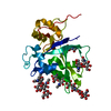
| ||||||||
|---|---|---|---|---|---|---|---|---|---|
| 1 |
| ||||||||
| Unit cell |
|
-Components
| #1: Protein | Mass: 22953.471 Da / Num. of mol.: 1 Source method: isolated from a genetically manipulated source Source: (gene. exp.) Gene: CERK1, RLK9, Os08g0538300, LOC_Os08g42580, P0665C04.34, P0666G10.101 Production host:  Insect BA phytoplasma (bacteria) Insect BA phytoplasma (bacteria)References: UniProt: A0A0P0XII1, non-specific serine/threonine protein kinase | ||||||
|---|---|---|---|---|---|---|---|
| #2: Polysaccharide | 2-acetamido-2-deoxy-beta-D-glucopyranose-(1-4)-2-acetamido-2-deoxy-beta-D-glucopyranose-(1-4)-2- ...2-acetamido-2-deoxy-beta-D-glucopyranose-(1-4)-2-acetamido-2-deoxy-beta-D-glucopyranose-(1-4)-2-acetamido-2-deoxy-beta-D-glucopyranose-(1-4)-2-acetamido-2-deoxy-beta-D-glucopyranose-(1-4)-2-acetamido-2-deoxy-beta-D-glucopyranose-(1-4)-2-acetamido-2-deoxy-beta-D-glucopyranose Source method: isolated from a genetically manipulated source | ||||||
| #3: Polysaccharide | beta-D-mannopyranose-(1-4)-2-acetamido-2-deoxy-beta-D-glucopyranose-(1-4)-[alpha-L-fucopyranose-(1- ...beta-D-mannopyranose-(1-4)-2-acetamido-2-deoxy-beta-D-glucopyranose-(1-4)-[alpha-L-fucopyranose-(1-3)][alpha-L-fucopyranose-(1-6)]2-acetamido-2-deoxy-beta-D-glucopyranose Type: oligosaccharide / Mass: 878.823 Da / Num. of mol.: 1 Source method: isolated from a genetically manipulated source | ||||||
| #4: Polysaccharide | Source method: isolated from a genetically manipulated source #5: Water | ChemComp-HOH / |  Mass: 18.015 Da / Num. of mol.: 38 / Source method: isolated from a natural source / Formula: H2O Mass: 18.015 Da / Num. of mol.: 38 / Source method: isolated from a natural source / Formula: H2OHas ligand of interest | Y | Has protein modification | Y | |
-Experimental details
-Experiment
| Experiment | Method: X-RAY DIFFRACTION / Number of used crystals: 1 |
|---|
- Sample preparation
| Crystal | Density Matthews: 2.62 Å3/Da / Density % sol: 53.02 % |
|---|---|
| Crystal grow | Temperature: 293 K / Method: vapor diffusion, hanging drop Details: 0.25M Magnesium chloride, 0.1 M TRIS, pH8.5, and 30% (w/v) PEG4000. |
-Data collection
| Diffraction | Mean temperature: 100 K / Serial crystal experiment: N |
|---|---|
| Diffraction source | Source: SYNCHROTRON / Site: SSRF  / Beamline: BL17U / Wavelength: 1 Å / Beamline: BL17U / Wavelength: 1 Å |
| Detector | Type: AGILENT EOS CCD / Detector: CCD / Date: May 1, 2018 |
| Radiation | Protocol: SINGLE WAVELENGTH / Monochromatic (M) / Laue (L): M / Scattering type: x-ray |
| Radiation wavelength | Wavelength: 1 Å / Relative weight: 1 |
| Reflection | Resolution: 2→50 Å / Num. obs: 15362 / % possible obs: 98.3 % / Redundancy: 5.4 % / Biso Wilson estimate: 31.59 Å2 / Rmerge(I) obs: 0.07 / Net I/σ(I): 25.78 |
| Reflection shell | Resolution: 2→2.07 Å / Rmerge(I) obs: 0.319 / Num. unique obs: 1486 |
- Processing
| Software |
| ||||||||||||||||||||||||||||||||||||||||||
|---|---|---|---|---|---|---|---|---|---|---|---|---|---|---|---|---|---|---|---|---|---|---|---|---|---|---|---|---|---|---|---|---|---|---|---|---|---|---|---|---|---|---|---|
| Refinement | Method to determine structure: MOLECULAR REPLACEMENT Starting model: 4EBZ Resolution: 2.015→42.175 Å / SU ML: 0.22 / Cross valid method: THROUGHOUT / σ(F): 1.35 / Phase error: 28.48 / Stereochemistry target values: ML
| ||||||||||||||||||||||||||||||||||||||||||
| Solvent computation | Shrinkage radii: 0.9 Å / VDW probe radii: 1.11 Å / Solvent model: FLAT BULK SOLVENT MODEL | ||||||||||||||||||||||||||||||||||||||||||
| Displacement parameters | Biso max: 95.5 Å2 / Biso mean: 42.8789 Å2 / Biso min: 20.93 Å2 | ||||||||||||||||||||||||||||||||||||||||||
| Refinement step | Cycle: final / Resolution: 2.015→42.175 Å
| ||||||||||||||||||||||||||||||||||||||||||
| Refine LS restraints |
| ||||||||||||||||||||||||||||||||||||||||||
| LS refinement shell | Refine-ID: X-RAY DIFFRACTION / Rfactor Rfree error: 0
| ||||||||||||||||||||||||||||||||||||||||||
| Refinement TLS params. | Method: refined / Origin x: 31.3453 Å / Origin y: -2.9306 Å / Origin z: 59.8769 Å
| ||||||||||||||||||||||||||||||||||||||||||
| Refinement TLS group | Selection details: (chain 'A' and resid 29 through 233) |
