 Movie
Movie Controller

[English] 日本語
 Yorodumi
- PDB-7vma: The X-ray crystallographic structure of amylo-alpha-1,6-glucosida... -
+ Open data

- Basic information
| Entry | Database: PDB / ID: 7vma | ||||||||||||
|---|---|---|---|---|---|---|---|---|---|---|---|---|---|
| Title | The X-ray crystallographic structure of amylo-alpha-1,6-glucosidase from Thermococcus gammatolerans STB12 | ||||||||||||
 Components Components | Amylo-alpha-1,6-glucosidase, putative archaeal type glycogen debranching enzyme (Gde) | ||||||||||||
 Keywords Keywords | HYDROLASE / Amylo-alpha-1 / 6-glucosidase | ||||||||||||
| Function / homology | Putative glycogen debranching enzyme, N-terminal / N-terminal domain of (some) glycogen debranching enzymes / amylo-alpha-1,6-glucosidase / amylo-alpha-1,6-glucosidase activity / Mannosylglycerate hydrolase MGH1-like glycoside hydrolase domain / Six-hairpin glycosidase-like superfamily / Six-hairpin glycosidase superfamily / carbohydrate metabolic process / Amylo-alpha-1,6-glucosidase, putative archaeal type glycogen debranching enzyme (Gde) Function and homology information Function and homology information | ||||||||||||
| Biological species |   Thermococcus gammatolerans EJ3 (archaea) Thermococcus gammatolerans EJ3 (archaea) | ||||||||||||
| Method |  X-RAY DIFFRACTION / SYNCHROTRON / MOLECULAR REPLACEMENT / Resolution: 2.804 Å X-RAY DIFFRACTION / SYNCHROTRON / MOLECULAR REPLACEMENT / Resolution: 2.804 Å | ||||||||||||
 Authors Authors | Li, Z.F. / Ban, X.F. / Wang, Y.M. / Li, C.M. / Gu, Z.B. | ||||||||||||
| Funding support |  China, 3items China, 3items
| ||||||||||||
 Citation Citation | Journal: To Be Published Title: The X-ray Crystallographic Structure of Amylo-alpha-1,6-glucosidase from Thermococcus gannatilerans STB12 Authors: Li, Z.F. / Ban, X.F. / Wang, Y.M. / Li, C.M. / Gu, Z.B. | ||||||||||||
| History |
|
- Structure visualization
| Structure viewer | Molecule: MolmilJmol/JSmol |
|---|
- Downloads & links
-Download
| PDBx/mmCIF format | 7vma.cif.gz | 245.6 KB | Display | PDBx/mmCIF format |
|---|---|---|---|---|
| PDB format | pdb7vma.ent.gz | 197.8 KB | Display | PDB format |
| PDBx/mmJSON format | 7vma.json.gz | Tree view | PDBx/mmJSON format | |
| Others |  Other downloads Other downloads |
-Validation report
| Summary document | 7vma_validation.pdf.gz | 449.9 KB | Display | wwPDB validaton report |
|---|---|---|---|---|
| Full document | 7vma_full_validation.pdf.gz | 494.2 KB | Display | |
| Data in XML | 7vma_validation.xml.gz | 48.3 KB | Display | |
| Data in CIF | 7vma_validation.cif.gz | 65.5 KB | Display | |
| Arichive directory | https://data.pdbj.org/pub/pdb/validation_reports/vm/7vmaftp://data.pdbj.org/pub/pdb/validation_reports/vm/7vma | HTTPS FTP |
-Related structure data
| Similar structure data |
|---|
-Links
 PDBj
PDBj
- Assembly
| Deposited unit | 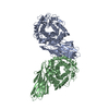
| ||||||||
|---|---|---|---|---|---|---|---|---|---|
| 1 | 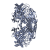
| ||||||||
| 2 | 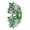
| ||||||||
| Unit cell |
|
-Components
| #1: Protein | Mass: 68705.500 Da / Num. of mol.: 2 Source method: isolated from a genetically manipulated source Source: (gene. exp.) Thermococcus gammatolerans EJ3 (archaea)Strain: DSM 15229 / JCM 11827 / EJ3 / Gene: gde, TGAM_1568 Production host:  References: UniProt: C5A758, amylo-alpha-1,6-glucosidase #2: Water | ChemComp-HOH / |  Mass: 18.015 Da / Num. of mol.: 14 / Source method: isolated from a natural source / Formula: H2O Mass: 18.015 Da / Num. of mol.: 14 / Source method: isolated from a natural source / Formula: H2O |
|---|
-Experimental details
-Experiment
| Experiment | Method: X-RAY DIFFRACTION / Number of used crystals: 1 |
|---|
- Sample preparation
| Crystal | Density Matthews: 2.4 Å3/Da / Density % sol: 48.71 % |
|---|---|
| Crystal grow | Temperature: 291 K / Method: vapor diffusion / Details: 25%tertiary butanol; 0.1M Tris |
-Data collection
| Diffraction | Mean temperature: 100 K / Serial crystal experiment: N |
|---|---|
| Diffraction source | Source: SYNCHROTRON / Site: SSRF / Beamline: BL19U1 / Wavelength: 0.9785 Å |
| Detector | Type: DECTRIS PILATUS3 6M / Detector: PIXEL / Date: May 7, 2021 |
| Radiation | Protocol: SINGLE WAVELENGTH / Monochromatic (M) / Laue (L): M / Scattering type: x-ray |
| Radiation wavelength | Wavelength: 0.9785 Å / Relative weight: 1 |
| Reflection | Resolution: 2.52→48.77 Å / Num. obs: 44230 / % possible obs: 99.8 % / Redundancy: 6.6 % / Rmerge(I) obs: 0.087 / Net I/σ(I): 12.3 |
| Reflection shell | Resolution: 2.52→2.6 Å / Rmerge(I) obs: 0.022 / Num. unique obs: 4569 |
- Processing
| Software |
| ||||||||||||||||||||||||||||||||||||||||||||||||||||||||||||||||||||||||||||||||||||
|---|---|---|---|---|---|---|---|---|---|---|---|---|---|---|---|---|---|---|---|---|---|---|---|---|---|---|---|---|---|---|---|---|---|---|---|---|---|---|---|---|---|---|---|---|---|---|---|---|---|---|---|---|---|---|---|---|---|---|---|---|---|---|---|---|---|---|---|---|---|---|---|---|---|---|---|---|---|---|---|---|---|---|---|---|---|
| Refinement | Method to determine structure: MOLECULAR REPLACEMENT Starting model: predict using alpha-fold Resolution: 2.804→38.998 Å / SU ML: 0.6 / Cross valid method: THROUGHOUT / σ(F): 1.34 / Phase error: 42.57 / Stereochemistry target values: ML
| ||||||||||||||||||||||||||||||||||||||||||||||||||||||||||||||||||||||||||||||||||||
| Solvent computation | Shrinkage radii: 0.9 Å / VDW probe radii: 1.11 Å / Solvent model: FLAT BULK SOLVENT MODEL | ||||||||||||||||||||||||||||||||||||||||||||||||||||||||||||||||||||||||||||||||||||
| Displacement parameters | Biso max: 150.94 Å2 / Biso mean: 94.3893 Å2 / Biso min: 57.17 Å2 | ||||||||||||||||||||||||||||||||||||||||||||||||||||||||||||||||||||||||||||||||||||
| Refinement step | Cycle: final / Resolution: 2.804→38.998 Å
| ||||||||||||||||||||||||||||||||||||||||||||||||||||||||||||||||||||||||||||||||||||
| LS refinement shell | Refine-ID: X-RAY DIFFRACTION / Rfactor Rfree error: 0 / Total num. of bins used: 11
|
