 Movie
Movie Controller


+ Open data

- Basic information
| Entry | Database: PDB / ID: 7vkp | ||||||
|---|---|---|---|---|---|---|---|
| Title | Crystal structure of E.coli pseudouridine kinase PsuK | ||||||
 Components Components | PfkB domain protein | ||||||
 Keywords Keywords | RNA BINDING PROTEIN / pseudouridine / kinase / yeiC | ||||||
| Function / homology |  Function and homology information Function and homology information | ||||||
| Biological species |  | ||||||
| Method |  X-RAY DIFFRACTION / SYNCHROTRON / MOLECULAR REPLACEMENT / Resolution: 2.3 Å X-RAY DIFFRACTION / SYNCHROTRON / MOLECULAR REPLACEMENT / Resolution: 2.3 Å | ||||||
 Authors Authors | Li, K.J. / Li, X.J. / Wu, B.X. | ||||||
| Funding support | 1items
| ||||||
 Citation Citation | Journal: Front Microbiol / Year: 2022 Title: Structure Characterization of Escherichia coli Pseudouridine Kinase PsuK. Authors: Li, X. / Li, K. / Guo, W. / Wen, Y. / Meng, C. / Wu, B. | ||||||
| History |
|
- Structure visualization
| Structure viewer | Molecule: MolmilJmol/JSmol |
|---|
- Downloads & links
-Download
| PDBx/mmCIF format | 7vkp.cif.gz | 154.9 KB | Display | PDBx/mmCIF format |
|---|---|---|---|---|
| PDB format | pdb7vkp.ent.gz | 102 KB | Display | PDB format |
| PDBx/mmJSON format | 7vkp.json.gz | Tree view | PDBx/mmJSON format | |
| Others |  Other downloads Other downloads |
-Validation report
| Arichive directory | https://data.pdbj.org/pub/pdb/validation_reports/vk/7vkpftp://data.pdbj.org/pub/pdb/validation_reports/vk/7vkp | HTTPS FTP |
|---|
-Related structure data
| Related structure data | 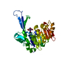 7vskC 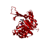 7w93C 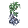 7c1yS S: Starting model for refinement C: citing same article ( |
|---|---|
| Similar structure data |
-Links
 PDBj
PDBj- Assembly
| Deposited unit | 
| ||||||||||||
|---|---|---|---|---|---|---|---|---|---|---|---|---|---|
| 1 |
| ||||||||||||
| Unit cell |
|
-Components
| #1: Protein | Mass: 33781.094 Da / Num. of mol.: 1 Source method: isolated from a genetically manipulated source Source: (gene. exp.) Strain: B / BL21-DE3 / Gene: ECBD_1492 / Production host: |
|---|---|
| #2: Water | ChemComp-HOH /  Mass: 18.015 Da / Num. of mol.: 37 / Source method: isolated from a natural source / Formula: H2O Mass: 18.015 Da / Num. of mol.: 37 / Source method: isolated from a natural source / Formula: H2O |
-Experimental details
-Experiment
| Experiment | Method: X-RAY DIFFRACTION / Number of used crystals: 1 |
|---|
- Sample preparation
| Crystal | Density Matthews: 3.87 Å3/Da / Density % sol: 68.22 % |
|---|---|
| Crystal grow | Temperature: 293 K / Method: vapor diffusion, sitting drop / pH: 7.5 Details: 0.02 M Magnesium chloride hexahydrate, 0.1 M HEPES pH 7.5, 22% w/v Poly(acrylic acid sodium salt) 5100 |
-Data collection
| Diffraction | Mean temperature: 100 K / Serial crystal experiment: N |
|---|---|
| Diffraction source | Source: SYNCHROTRON / Site: SSRF  / Beamline: BL18U1 / Wavelength: 0.97915 Å / Beamline: BL18U1 / Wavelength: 0.97915 Å |
| Detector | Type: DECTRIS PILATUS 6M / Detector: PIXEL / Date: Sep 21, 2021 |
| Radiation | Protocol: SINGLE WAVELENGTH / Monochromatic (M) / Laue (L): M / Scattering type: x-ray |
| Radiation wavelength | Wavelength: 0.97915 Å / Relative weight: 1 |
| Reflection | Resolution: 2.3→30 Å / Num. obs: 24045 / % possible obs: 99.9 % / Redundancy: 21 % / Biso Wilson estimate: 23.14 Å2 / CC1/2: 1 / Rmerge(I) obs: 0.148 / Rpim(I) all: 0.029 / Rrim(I) all: 0.151 / Net I/σ(I): 22.25 |
| Reflection shell | Resolution: 2.3→2.38 Å / Redundancy: 9.6 % / Rmerge(I) obs: 0.52 / Mean I/σ(I) obs: 3.5 / Num. unique obs: 2362 / CC1/2: 0.294 / CC star: 0.674 / Rpim(I) all: 0.169 / Rrim(I) all: 0.549 / % possible all: 99.9 |
- Processing
| Software |
| |||||||||||||||||||||||||||||||||||||||||||||||||||||||||||||||
|---|---|---|---|---|---|---|---|---|---|---|---|---|---|---|---|---|---|---|---|---|---|---|---|---|---|---|---|---|---|---|---|---|---|---|---|---|---|---|---|---|---|---|---|---|---|---|---|---|---|---|---|---|---|---|---|---|---|---|---|---|---|---|---|---|
| Refinement | Method to determine structure: MOLECULAR REPLACEMENT Starting model: 7C1Y Resolution: 2.3→29.18 Å / SU ML: 0.2796 / Cross valid method: FREE R-VALUE / σ(F): 1.53 / Phase error: 23.7643 Stereochemistry target values: GeoStd + Monomer Library + CDL v1.2
| |||||||||||||||||||||||||||||||||||||||||||||||||||||||||||||||
| Solvent computation | Shrinkage radii: 0.9 Å / VDW probe radii: 1.11 Å / Solvent model: FLAT BULK SOLVENT MODEL | |||||||||||||||||||||||||||||||||||||||||||||||||||||||||||||||
| Displacement parameters | Biso mean: 42.24 Å2 | |||||||||||||||||||||||||||||||||||||||||||||||||||||||||||||||
| Refinement step | Cycle: LAST / Resolution: 2.3→29.18 Å
| |||||||||||||||||||||||||||||||||||||||||||||||||||||||||||||||
| Refine LS restraints |
| |||||||||||||||||||||||||||||||||||||||||||||||||||||||||||||||
| LS refinement shell |
| |||||||||||||||||||||||||||||||||||||||||||||||||||||||||||||||
| Refinement TLS params. | Method: refined / Origin x: 74.4502827355 Å / Origin y: -20.2127539345 Å / Origin z: -3.90177952866 Å
| |||||||||||||||||||||||||||||||||||||||||||||||||||||||||||||||
| Refinement TLS group | Selection details: all |
