 Movie
Movie Controller


+ Open data

- Basic information
| Entry | Database: PDB / ID: 7vkb | ||||||
|---|---|---|---|---|---|---|---|
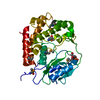
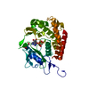
| Title | Crystal structure of the a bacterial kinase complex | ||||||
 Components Components |
| ||||||
 Keywords Keywords | TOXIN/ANTITOXIN / TOXIN ANTITOXIN SYSTEM / TOXIN / TOXIN-ANTITOXIN complex | ||||||
| Function / homology |  Function and homology information Function and homology information | ||||||
| Biological species |  Legionella pneumophila subsp. pneumophila str. Philadelphia 1 (bacteria) Legionella pneumophila subsp. pneumophila str. Philadelphia 1 (bacteria) | ||||||
| Method |  X-RAY DIFFRACTION / SYNCHROTRON / SAD / Resolution: 1.82 Å X-RAY DIFFRACTION / SYNCHROTRON / SAD / Resolution: 1.82 Å | ||||||
 Authors Authors | Zhen, X. / Ouyang, S.Y. | ||||||
| Funding support |  China, 1items China, 1items
| ||||||
 Citation Citation | Journal: Nat Commun / Year: 2022 Title: Molecular mechanism of toxin neutralization in the HipBST toxin-antitoxin system of Legionella pneumophila. Authors: Zhen, X. / Wu, Y. / Ge, J. / Fu, J. / Ye, L. / Lin, N. / Huang, Z. / Liu, Z. / Luo, Z.Q. / Qiu, J. / Ouyang, S. | ||||||
| History |
|
- Structure visualization
| Structure viewer | Molecule: MolmilJmol/JSmol |
|---|
- Downloads & links
-Download
| PDBx/mmCIF format | 7vkb.cif.gz | 167.6 KB | Display | PDBx/mmCIF format |
|---|---|---|---|---|
| PDB format | pdb7vkb.ent.gz | 130.8 KB | Display | PDB format |
| PDBx/mmJSON format | 7vkb.json.gz | Tree view | PDBx/mmJSON format | |
| Others |  Other downloads Other downloads |
-Validation report
| Arichive directory | https://data.pdbj.org/pub/pdb/validation_reports/vk/7vkbftp://data.pdbj.org/pub/pdb/validation_reports/vk/7vkb | HTTPS FTP |
|---|
-Related structure data
-Links
 PDBj
PDBj- Assembly
| Deposited unit | 
| ||||||||
|---|---|---|---|---|---|---|---|---|---|
| 1 |
| ||||||||
| Unit cell |
| ||||||||
| Components on special symmetry positions |
|
-Components
| #1: Protein | Mass: 36957.707 Da / Num. of mol.: 1 Source method: isolated from a genetically manipulated source Source: (gene. exp.) Legionella pneumophila subsp. pneumophila str. Philadelphia 1 (bacteria)Gene: lpg2370 Production host: References: UniProt: Q5ZSZ6 |
|---|---|
| #2: Protein | Mass: 11709.013 Da / Num. of mol.: 1 Source method: isolated from a genetically manipulated source Source: (gene. exp.) Legionella pneumophila subsp. pneumophila str. Philadelphia 1 (bacteria)Gene: lpg2369 / Production host: |
| #3: Chemical | ChemComp-NA /   Mass: 22.990 Da / Num. of mol.: 1 / Source method: obtained synthetically / Formula: Na / Feature type: SUBJECT OF INVESTIGATION Mass: 22.990 Da / Num. of mol.: 1 / Source method: obtained synthetically / Formula: Na / Feature type: SUBJECT OF INVESTIGATION |
| #4: Chemical | ChemComp-PEG /   Mass: 106.120 Da / Num. of mol.: 1 / Source method: obtained synthetically / Formula: C4H10O3 / Feature type: SUBJECT OF INVESTIGATION Mass: 106.120 Da / Num. of mol.: 1 / Source method: obtained synthetically / Formula: C4H10O3 / Feature type: SUBJECT OF INVESTIGATION |
| #5: Water | ChemComp-HOH /  Mass: 18.015 Da / Num. of mol.: 243 / Source method: isolated from a natural source / Formula: H2O Mass: 18.015 Da / Num. of mol.: 243 / Source method: isolated from a natural source / Formula: H2O |
| Has ligand of interest | Y |
| Has protein modification | Y |
-Experimental details
-Experiment
| Experiment | Method: X-RAY DIFFRACTION / Number of used crystals: 1 |
|---|
- Sample preparation
| Crystal | Density Matthews: 2.07 Å3/Da / Density % sol: 40.64 % |
|---|---|
| Crystal grow | Temperature: 289 K / Method: vapor diffusion, hanging drop / pH: 7.2 / Details: 0.1M Sodium acetate PH 7.0 12% PEG 3350 / PH range: 6-8 |
-Data collection
| Diffraction | Mean temperature: 100 K / Serial crystal experiment: N |
|---|---|
| Diffraction source | Source: SYNCHROTRON / Site: SSRF / Beamline: BL17U1 / Wavelength: 0.97981 Å |
| Detector | Type: ADSC QUANTUM 315r / Detector: CCD / Date: Dec 30, 2020 |
| Radiation | Protocol: SINGLE WAVELENGTH / Monochromatic (M) / Laue (L): M / Scattering type: x-ray |
| Radiation wavelength | Wavelength: 0.97981 Å / Relative weight: 1 |
| Reflection | Resolution: 1.82→31.871 Å / Num. obs: 38842 / % possible obs: 100 % / Redundancy: 15.1 % / Biso Wilson estimate: 38.79 Å2 / CC1/2: 1 / Net I/σ(I): 2 |
| Reflection shell | Resolution: 1.82→1.87 Å / Rmerge(I) obs: 0.089 / Num. unique obs: 2774 / CC1/2: 0.801 / Rpim(I) all: 0.091 / % possible all: 100 |
- Processing
| Software |
| ||||||||||||||||||||||||||||||||||||||||||||||||||||||||||||||||||||||||||||||||||||||||||
|---|---|---|---|---|---|---|---|---|---|---|---|---|---|---|---|---|---|---|---|---|---|---|---|---|---|---|---|---|---|---|---|---|---|---|---|---|---|---|---|---|---|---|---|---|---|---|---|---|---|---|---|---|---|---|---|---|---|---|---|---|---|---|---|---|---|---|---|---|---|---|---|---|---|---|---|---|---|---|---|---|---|---|---|---|---|---|---|---|---|---|---|
| Refinement | Method to determine structure: SAD / Resolution: 1.82→31.87 Å / SU ML: 0.19 / Cross valid method: THROUGHOUT / σ(F): 1.34 / Phase error: 23.51 / Stereochemistry target values: ML
| ||||||||||||||||||||||||||||||||||||||||||||||||||||||||||||||||||||||||||||||||||||||||||
| Solvent computation | Shrinkage radii: 0.9 Å / VDW probe radii: 1.1 Å / Solvent model: FLAT BULK SOLVENT MODEL | ||||||||||||||||||||||||||||||||||||||||||||||||||||||||||||||||||||||||||||||||||||||||||
| Displacement parameters | Biso max: 160.45 Å2 / Biso mean: 46.2556 Å2 / Biso min: 27.28 Å2 | ||||||||||||||||||||||||||||||||||||||||||||||||||||||||||||||||||||||||||||||||||||||||||
| Refinement step | Cycle: final / Resolution: 1.82→31.87 Å
| ||||||||||||||||||||||||||||||||||||||||||||||||||||||||||||||||||||||||||||||||||||||||||
| LS refinement shell | Refine-ID: X-RAY DIFFRACTION / Rfactor Rfree error: 0 / Total num. of bins used: 14 / % reflection obs: 100 %
| ||||||||||||||||||||||||||||||||||||||||||||||||||||||||||||||||||||||||||||||||||||||||||
| Refinement TLS params. | Method: refined / Origin x: 25.8055 Å / Origin y: 18.4674 Å / Origin z: 73.6968 Å
| ||||||||||||||||||||||||||||||||||||||||||||||||||||||||||||||||||||||||||||||||||||||||||
| Refinement TLS group |
|
