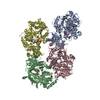
| Deposited unit | D: D-alanine--D-alanyl carrier protein ligase
C: D-alanine--D-alanyl carrier protein ligase
A: D-alanine--D-alanyl carrier protein ligase
B: D-alanine--D-alanyl carrier protein ligase
hetero molecules
| Theoretical mass | Number of molelcules |
|---|
| Total (without water) | 220,907 | 12 |
|---|
| Polymers | 218,781 | 4 |
|---|
| Non-polymers | 2,126 | 8 |
|---|
| Water | 6,864 | 381 |
|---|
|
|---|
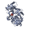
| 1 | C: D-alanine--D-alanyl carrier protein ligase
hetero molecules
| Theoretical mass | Number of molelcules |
|---|
| Total (without water) | 55,227 | 3 |
|---|
| Polymers | 54,695 | 1 |
|---|
| Non-polymers | 531 | 2 |
|---|
| Water | 18 | 1 |
|---|
| Type | Name | Symmetry operation | Number |
|---|
| identity operation | 1_555 | x,y,z | 1 |
| Buried area | 1070 Å2 |
|---|
| ΔGint | -12 kcal/mol |
|---|
| Surface area | 20180 Å2 |
|---|
| Method | PISA |
|---|
|
|---|
| 2 | D: D-alanine--D-alanyl carrier protein ligase
hetero molecules
| Theoretical mass | Number of molelcules |
|---|
| Total (without water) | 55,227 | 3 |
|---|
| Polymers | 54,695 | 1 |
|---|
| Non-polymers | 531 | 2 |
|---|
| Water | 18 | 1 |
|---|
| Type | Name | Symmetry operation | Number |
|---|
| identity operation | 1_555 | x,y,z | 1 |
| Buried area | 1000 Å2 |
|---|
| ΔGint | -12 kcal/mol |
|---|
| Surface area | 20720 Å2 |
|---|
| Method | PISA |
|---|
|
|---|
| 3 | B: D-alanine--D-alanyl carrier protein ligase
hetero molecules
| Theoretical mass | Number of molelcules |
|---|
| Total (without water) | 55,227 | 3 |
|---|
| Polymers | 54,695 | 1 |
|---|
| Non-polymers | 531 | 2 |
|---|
| Water | 18 | 1 |
|---|
| Type | Name | Symmetry operation | Number |
|---|
| identity operation | 1_555 | x,y,z | 1 |
| Buried area | 1080 Å2 |
|---|
| ΔGint | -12 kcal/mol |
|---|
| Surface area | 20080 Å2 |
|---|
| Method | PISA |
|---|
|
|---|
| 4 | A: D-alanine--D-alanyl carrier protein ligase
hetero molecules
| Theoretical mass | Number of molelcules |
|---|
| Total (without water) | 55,227 | 3 |
|---|
| Polymers | 54,695 | 1 |
|---|
| Non-polymers | 531 | 2 |
|---|
| Water | 18 | 1 |
|---|
| Type | Name | Symmetry operation | Number |
|---|
| identity operation | 1_555 | x,y,z | 1 |
| Buried area | 1080 Å2 |
|---|
| ΔGint | -12 kcal/mol |
|---|
| Surface area | 20250 Å2 |
|---|
| Method | PISA |
|---|
|
|---|
| Unit cell | | Length a, b, c (Å) | 89.468, 88.510, 130.850 |
|---|
| Angle α, β, γ (deg.) | 90.000, 91.670, 90.000 |
|---|
| Int Tables number | 4 |
|---|
| Space group name H-M | P1211 |
|---|
|
|---|
| Noncrystallographic symmetry (NCS) | NCS domain: | ID | Ens-ID | Details |
|---|
| 1 | 1 | (chain A and (resseq 2 or (resid 3 and (name...| 2 | 1 | (chain B and (resseq 2 or (resid 3 and (name...| 3 | 1 | (chain C and (resseq 2 or (resid 3 and (name...| 4 | 1 | (chain D and (resseq 2 or (resid 3 and (name... | | | |
NCS domain segments: | Dom-ID | Component-ID | Ens-ID | Selection details | Auth asym-ID | Auth seq-ID |
|---|
| 1 | 1 | 1 | (chain A and (resseq 2 or (resid 3 and (name...A| 2 | | 1 | 2 | 1 | (chain A and (resseq 2 or (resid 3 and (name...A| 3 | | 1 | 3 | 1 | (chain A and (resseq 2 or (resid 3 and (name...A| 1 - 511 | | 1 | 4 | 1 | (chain A and (resseq 2 or (resid 3 and (name...A| 1 - 511 | | 1 | 5 | 1 | (chain A and (resseq 2 or (resid 3 and (name...A| 1 - 511 | | 1 | 6 | 1 | (chain A and (resseq 2 or (resid 3 and (name...A| 1 - 511 | | 1 | 7 | 1 | (chain A and (resseq 2 or (resid 3 and (name...A| 1 - 511 | | 1 | 8 | 1 | (chain A and (resseq 2 or (resid 3 and (name...A| 1 - 511 | | 2 | 1 | 1 | (chain B and (resseq 2 or (resid 3 and (name...B| 2 | | 2 | 2 | 1 | (chain B and (resseq 2 or (resid 3 and (name | | | | | | | | | | | | | | | | | | |
|
|---|
 Movie
Movie Controller

 Yorodumi
 Open data
 Basic information
 Components
 Keywords
 Function and homology information
 Staphylococcus aureus subsp. aureus Mu50 (bacteria)
 X-RAY DIFFRACTION /
  Authors
 Korea, Republic Of, 2items
  Citation
 Structure visualization
 Downloads & links
 Other downloads
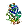
 PDBj
PDBj
 Assembly