Movie
Movie Controller

[English] 日本語
 Yorodumi
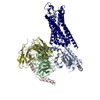
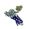
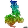
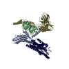
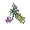
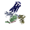
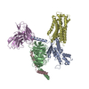
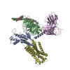
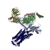
- PDB-7vgx: Neuropeptide Y Y1 Receptor (NPY1R) in Complex with G Protein and ... -
+ Open data

- Basic information
| Entry | Database: PDB / ID: 7vgx | |||||||||
|---|---|---|---|---|---|---|---|---|---|---|
| Title | Neuropeptide Y Y1 Receptor (NPY1R) in Complex with G Protein and its endogeneous Peptide-Agonist Neuropeptide Y (NPY) | |||||||||
 Components Components |
| |||||||||
 Keywords Keywords | MEMBRANE PROTEIN / NPY / GPCR / G-protein / Complex | |||||||||
| Function / homology |  Function and homology information Function and homology informationpancreatic polypeptide receptor activity / peptide YY receptor activity / neuropeptide Y receptor activity / synaptic signaling via neuropeptide / neuropeptide Y receptor binding / intestinal epithelial cell differentiation / positive regulation of appetite / neuropeptide receptor activity / adult feeding behavior / neuropeptide hormone activity ...pancreatic polypeptide receptor activity / peptide YY receptor activity / neuropeptide Y receptor activity / synaptic signaling via neuropeptide / neuropeptide Y receptor binding / intestinal epithelial cell differentiation / positive regulation of appetite / neuropeptide receptor activity / adult feeding behavior / neuropeptide hormone activity / neuropeptide binding / feeding behavior / central nervous system neuron development / outflow tract morphogenesis / neuronal dense core vesicle / G protein-coupled receptor signaling pathway, coupled to cyclic nucleotide second messenger / regulation of multicellular organism growth / calcium channel regulator activity / GABA-ergic synapse / Adenylate cyclase inhibitory pathway / neuropeptide signaling pathway / positive regulation of protein localization to cell cortex / regulation of cAMP-mediated signaling / FOXO-mediated transcription of oxidative stress, metabolic and neuronal genes / D2 dopamine receptor binding / G protein-coupled serotonin receptor binding / regulation of mitotic spindle organization / cellular response to forskolin / sensory perception of pain / adenylate cyclase-inhibiting G protein-coupled receptor signaling pathway / Peptide ligand-binding receptors / locomotory behavior / Regulation of insulin secretion / G protein-coupled receptor binding / G protein-coupled receptor activity / G-protein beta/gamma-subunit complex binding / Olfactory Signaling Pathway / Activation of the phototransduction cascade / adenylate cyclase-modulating G protein-coupled receptor signaling pathway / G beta:gamma signalling through PLC beta / Presynaptic function of Kainate receptors / Thromboxane signalling through TP receptor / G-protein activation / G protein-coupled acetylcholine receptor signaling pathway / Activation of G protein gated Potassium channels / Inhibition of voltage gated Ca2+ channels via Gbeta/gamma subunits / Prostacyclin signalling through prostacyclin receptor / Glucagon signaling in metabolic regulation / G beta:gamma signalling through CDC42 / cerebral cortex development / ADP signalling through P2Y purinoceptor 12 / G beta:gamma signalling through BTK / response to peptide hormone / Synthesis, secretion, and inactivation of Glucagon-like Peptide-1 (GLP-1) / regulation of blood pressure / Sensory perception of sweet, bitter, and umami (glutamate) taste / photoreceptor disc membrane / Adrenaline,noradrenaline inhibits insulin secretion / Glucagon-type ligand receptors / Vasopressin regulates renal water homeostasis via Aquaporins / G alpha (z) signalling events / cellular response to catecholamine stimulus / Glucagon-like Peptide-1 (GLP1) regulates insulin secretion / ADORA2B mediated anti-inflammatory cytokines production / sensory perception of taste / ADP signalling through P2Y purinoceptor 1 / adenylate cyclase-activating dopamine receptor signaling pathway / G beta:gamma signalling through PI3Kgamma / cellular response to prostaglandin E stimulus / Cooperation of PDCL (PhLP1) and TRiC/CCT in G-protein beta folding / glucose metabolic process / GPER1 signaling / GDP binding / G-protein beta-subunit binding / Inactivation, recovery and regulation of the phototransduction cascade / heterotrimeric G-protein complex / neuron projection development / G alpha (12/13) signalling events / extracellular vesicle / signaling receptor complex adaptor activity / Thrombin signalling through proteinase activated receptors (PARs) / GTPase binding / retina development in camera-type eye / phospholipase C-activating G protein-coupled receptor signaling pathway / Ca2+ pathway / cell cortex / midbody / G alpha (i) signalling events / fibroblast proliferation / G alpha (s) signalling events / G alpha (q) signalling events / chemical synaptic transmission / cell population proliferation / Ras protein signal transduction / Extra-nuclear estrogen signaling / neuron projection / cell cycle / G protein-coupled receptor signaling pathway / cell division / lysosomal membrane Similarity search - Function | |||||||||
| Biological species |  Homo sapiens (human) Homo sapiens (human) | |||||||||
| Method | ELECTRON MICROSCOPY / single particle reconstruction / cryo EM / Resolution: 3.2 Å | |||||||||
 Authors Authors | Park, C. / Kim, J. / Jeong, H. / Kang, H. / Bang, I. / Choi, H.-J. | |||||||||
| Funding support |  Korea, Republic Of, 2items Korea, Republic Of, 2items
| |||||||||
 Citation Citation | Journal: Nat Commun / Year: 2022 Title: Structural basis of neuropeptide Y signaling through Y1 receptor. Authors: Chaehee Park / Jinuk Kim / Seung-Bum Ko / Yeol Kyo Choi / Hyeongseop Jeong / Hyeonuk Woo / Hyunook Kang / Injin Bang / Sang Ah Kim / Tae-Young Yoon / Chaok Seok / Wonpil Im / Hee-Jung Choi /  Abstract: Neuropeptide Y (NPY) is highly abundant in the brain and involved in various physiological processes related to food intake and anxiety, as well as human diseases such as obesity and cancer. However, ...Neuropeptide Y (NPY) is highly abundant in the brain and involved in various physiological processes related to food intake and anxiety, as well as human diseases such as obesity and cancer. However, the molecular details of the interactions between NPY and its receptors are poorly understood. Here, we report a cryo-electron microscopy structure of the NPY-bound neuropeptide Y1 receptor (YR) in complex with G protein. The NPY C-terminal segment forming the extended conformation binds deep into the YR transmembrane core, where the amidated C-terminal residue Y36 of NPY is located at the base of the ligand-binding pocket. Furthermore, the helical region and two N-terminal residues of NPY interact with YR extracellular loops, contributing to the high affinity of NPY for YR. The structural analysis of NPY-bound YR and mutagenesis studies provide molecular insights into the activation mechanism of YR upon NPY binding. | |||||||||
| History |
|
- Structure visualization
| Movie |
Movie viewer |
|---|---|
| Structure viewer | Molecule: MolmilJmol/JSmol |
- Downloads & links
-Download
| PDBx/mmCIF format | 7vgx.cif.gz | 234.6 KB | Display | PDBx/mmCIF format |
|---|---|---|---|---|
| PDB format | pdb7vgx.ent.gz | 184.6 KB | Display | PDB format |
| PDBx/mmJSON format | 7vgx.json.gz | Tree view | PDBx/mmJSON format | |
| Others |  Other downloads Other downloads |
-Validation report
| Summary document | 7vgx_validation.pdf.gz | 749.1 KB | Display | wwPDB validaton report |
|---|---|---|---|---|
| Full document | 7vgx_full_validation.pdf.gz | 758.1 KB | Display | |
| Data in XML | 7vgx_validation.xml.gz | 36 KB | Display | |
| Data in CIF | 7vgx_validation.cif.gz | 54.9 KB | Display | |
| Arichive directory | https://data.pdbj.org/pub/pdb/validation_reports/vg/7vgxftp://data.pdbj.org/pub/pdb/validation_reports/vg/7vgx | HTTPS FTP |
-Related structure data
| Related structure data |  31979MC M: map data used to model this data C: citing same article ( |
|---|---|
| Similar structure data |
-Links
 PDBj
PDBj


- Assembly
| Deposited unit | 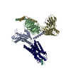
|
|---|---|
| 1 |
|
-Components
-Neuropeptide ... , 2 types, 2 molecules RL
| #1: Protein | Mass: 46489.961 Da / Num. of mol.: 1 Source method: isolated from a genetically manipulated source Source: (gene. exp.) Homo sapiens (human) / Gene: NPY1R, NPYR, NPYY1 / Cell line (production host): Sf9 / Production host:   Spodoptera frugiperda (fall armyworm) / References: UniProt: P25929 Spodoptera frugiperda (fall armyworm) / References: UniProt: P25929 |
|---|---|
| #2: Protein/peptide | Mass: 4277.735 Da / Num. of mol.: 1 / Source method: obtained synthetically / Source: (synth.) Homo sapiens (human) / References: UniProt: P01303 |
-Guanine nucleotide-binding protein ... , 3 types, 3 molecules ABG
| #3: Protein | Mass: 40427.969 Da / Num. of mol.: 1 Source method: isolated from a genetically manipulated source Source: (gene. exp.) Homo sapiens (human) / Gene: GNAI1 / Production host:  |
|---|---|
| #4: Protein | Mass: 37728.152 Da / Num. of mol.: 1 Source method: isolated from a genetically manipulated source Source: (gene. exp.) Homo sapiens (human) / Gene: GNB1 / Cell line (production host): Sf9 / Production host: Spodoptera frugiperda (fall armyworm) / References: UniProt: P62873 |
| #5: Protein | Mass: 7845.078 Da / Num. of mol.: 1 / Mutation: C68S Source method: isolated from a genetically manipulated source Source: (gene. exp.) Homo sapiens (human) / Gene: GNG2 / Production host: Spodoptera frugiperda (fall armyworm) / References: UniProt: P59768 |
-Antibody , 1 types, 1 molecules S
| #6: Antibody | Mass: 27784.896 Da / Num. of mol.: 1 Source method: isolated from a genetically manipulated source Source: (gene. exp.) Trichoplusia ni (cabbage looper) |
|---|
-Details
| Has ligand of interest | Y |
|---|---|
| Sequence details | Last 11 residues (AAAHHHHHHH |
-Experimental details
-Experiment
| Experiment | Method: ELECTRON MICROSCOPY |
|---|---|
| EM experiment | Aggregation state: PARTICLE / 3D reconstruction method: single particle reconstruction |
- Sample preparation
| Component |
| ||||||||||||||||||||||||||||||||||||
|---|---|---|---|---|---|---|---|---|---|---|---|---|---|---|---|---|---|---|---|---|---|---|---|---|---|---|---|---|---|---|---|---|---|---|---|---|---|
| Molecular weight | Value: 0.16 MDa / Experimental value: NO | ||||||||||||||||||||||||||||||||||||
| Source (natural) |
| ||||||||||||||||||||||||||||||||||||
| Source (recombinant) |
| ||||||||||||||||||||||||||||||||||||
| Buffer solution | pH: 8 | ||||||||||||||||||||||||||||||||||||
| Buffer component |
| ||||||||||||||||||||||||||||||||||||
| Specimen | Conc.: 10 mg/ml / Embedding applied: NO / Shadowing applied: NO / Staining applied: NO / Vitrification applied: YES | ||||||||||||||||||||||||||||||||||||
| Specimen support | Grid material: COPPER / Grid mesh size: 300 divisions/in. / Grid type: Quantifoil R1.2/1.3 | ||||||||||||||||||||||||||||||||||||
| Vitrification | Instrument: FEI VITROBOT MARK IV / Cryogen name: ETHANE / Humidity: 100 % / Chamber temperature: 285 K / Details: blot for 5 seconds before plunging |
- Electron microscopy imaging
| Experimental equipment |  Model: Titan Krios / Image courtesy: FEI Company |
|---|---|
| Microscopy | Model: FEI TITAN KRIOS |
| Electron gun | Electron source:  FIELD EMISSION GUN / Accelerating voltage: 300 kV / Illumination mode: FLOOD BEAM FIELD EMISSION GUN / Accelerating voltage: 300 kV / Illumination mode: FLOOD BEAM |
| Electron lens | Mode: BRIGHT FIELD / Nominal magnification: 75000 X / Nominal defocus max: 3750 nm / Nominal defocus min: 1250 nm / Cs: 0.1 mm |
| Specimen holder | Cryogen: NITROGEN |
| Image recording | Electron dose: 40 e/Å2 / Detector mode: COUNTING / Film or detector model: FEI FALCON III (4k x 4k) |
| EM imaging optics | Spherical aberration corrector: Microscope was modified with a Cs corrector |
| Image scans | Width: 4096 / Height: 4096 |
- Processing
| Software |
| ||||||||||||||||||||||||||||||||
|---|---|---|---|---|---|---|---|---|---|---|---|---|---|---|---|---|---|---|---|---|---|---|---|---|---|---|---|---|---|---|---|---|---|
| EM software |
| ||||||||||||||||||||||||||||||||
| CTF correction | Type: PHASE FLIPPING AND AMPLITUDE CORRECTION | ||||||||||||||||||||||||||||||||
| Particle selection | Num. of particles selected: 1339224 | ||||||||||||||||||||||||||||||||
| Symmetry | Point symmetry: C1 (asymmetric) | ||||||||||||||||||||||||||||||||
| 3D reconstruction | Resolution: 3.2 Å / Resolution method: FSC 0.143 CUT-OFF / Num. of particles: 233117 / Symmetry type: POINT | ||||||||||||||||||||||||||||||||
| Atomic model building | Protocol: RIGID BODY FIT / Space: REAL | ||||||||||||||||||||||||||||||||
| Atomic model building |
| ||||||||||||||||||||||||||||||||
| Refinement | Cross valid method: NONE Stereochemistry target values: GeoStd + Monomer Library + CDL v1.2 | ||||||||||||||||||||||||||||||||
| Displacement parameters | Biso mean: 71.54 Å2 | ||||||||||||||||||||||||||||||||
| Refine LS restraints |
|