 Movie
Movie Controller

[English] 日本語
 Yorodumi
- PDB-7vfr: GltA N83K mutant from Bifidobacterium infantis JCM 1222 complexed... -
+ Open data

- Basic information
| Entry | Database: PDB / ID: 7vfr | ||||||||||||
|---|---|---|---|---|---|---|---|---|---|---|---|---|---|
| Title | GltA N83K mutant from Bifidobacterium infantis JCM 1222 complexed with lacto-N-tetraose | ||||||||||||
 Components Components | Extracellular solute-binding protein, family 1 | ||||||||||||
 Keywords Keywords | TRANSLOCASE / solute-binding protein | ||||||||||||
| Function / homology | : / Bacterial extracellular solute-binding protein / Bacterial extracellular solute-binding protein / Prokaryotic membrane lipoprotein lipid attachment site profile. / metal ion binding / Extracellular solute-binding protein, family 1 Function and homology information Function and homology information | ||||||||||||
| Biological species |  Bifidobacterium longum subsp. infantis (bacteria) Bifidobacterium longum subsp. infantis (bacteria) | ||||||||||||
| Method |  X-RAY DIFFRACTION / SYNCHROTRON / MOLECULAR REPLACEMENT / Resolution: 1.56 Å X-RAY DIFFRACTION / SYNCHROTRON / MOLECULAR REPLACEMENT / Resolution: 1.56 Å | ||||||||||||
 Authors Authors | Sato, M. / Sakanaka, M. / Katayama, T. / Fushinobu, S. | ||||||||||||
| Funding support |  Japan, 3items Japan, 3items
| ||||||||||||
 Citation Citation | Journal: To Be Published Title: Short stretch sequence of a milk oligosaccharide transporter represents the phenotypic selection trajectory in infant gut microbiome Authors: Sakanaka, M. / Sato, M. / Fushinobu, S. / Katayama, T. | ||||||||||||
| History |
|
- Structure visualization
| Structure viewer | Molecule: MolmilJmol/JSmol |
|---|
- Downloads & links
-Download
| PDBx/mmCIF format | 7vfr.cif.gz | 103.6 KB | Display | PDBx/mmCIF format |
|---|---|---|---|---|
| PDB format | pdb7vfr.ent.gz | 73.4 KB | Display | PDB format |
| PDBx/mmJSON format | 7vfr.json.gz | Tree view | PDBx/mmJSON format | |
| Others |  Other downloads Other downloads |
-Validation report
| Arichive directory | https://data.pdbj.org/pub/pdb/validation_reports/vf/7vfrftp://data.pdbj.org/pub/pdb/validation_reports/vf/7vfr | HTTPS FTP |
|---|
-Related structure data
| Related structure data |  7vfqC  2z8fS S: Starting model for refinement C: citing same article ( |
|---|---|
| Similar structure data |
-Links
 PDBj
PDBj


- Assembly
| Deposited unit | 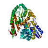
| ||||||||
|---|---|---|---|---|---|---|---|---|---|
| 1 |
| ||||||||
| Unit cell |
|
-Components
| #1: Protein | Mass: 43845.848 Da / Num. of mol.: 1 / Mutation: N83K Source method: isolated from a genetically manipulated source Source: (gene. exp.) Bifidobacterium longum subsp. infantis (strain ATCC 15697 / DSM 20088 / JCM 1222 / NCTC 11817 / S12) (bacteria)Gene: Blon_2177 / Plasmid: pET23b / Production host: | ||||||
|---|---|---|---|---|---|---|---|
| #2: Polysaccharide | beta-D-galactopyranose-(1-3)-2-acetamido-2-deoxy-beta-D-glucopyranose-(1-3)-beta-D-galactopyranose- ...beta-D-galactopyranose-(1-3)-2-acetamido-2-deoxy-beta-D-glucopyranose-(1-3)-beta-D-galactopyranose-(1-4)-beta-D-glucopyranose Source method: isolated from a genetically manipulated source | ||||||
| #3: Chemical |   Mass: 96.063 Da / Num. of mol.: 2 / Source method: obtained synthetically / Formula: SO4 Mass: 96.063 Da / Num. of mol.: 2 / Source method: obtained synthetically / Formula: SO4#4: Chemical | ChemComp-ZN / |   Mass: 65.409 Da / Num. of mol.: 1 / Source method: obtained synthetically / Formula: Zn Mass: 65.409 Da / Num. of mol.: 1 / Source method: obtained synthetically / Formula: Zn#5: Water | ChemComp-HOH / |  Mass: 18.015 Da / Num. of mol.: 397 / Source method: isolated from a natural source / Formula: H2O Mass: 18.015 Da / Num. of mol.: 397 / Source method: isolated from a natural source / Formula: H2OHas ligand of interest | Y | |
-Experimental details
-Experiment
| Experiment | Method: X-RAY DIFFRACTION / Number of used crystals: 1 |
|---|
- Sample preparation
| Crystal | Density Matthews: 2.03 Å3/Da / Density % sol: 39.54 % |
|---|---|
| Crystal grow | Temperature: 293.15 K / Method: vapor diffusion, sitting drop / pH: 5.5 Details: 30% (w/v) PEG MME 550, 10mM ZnSO4, 0.1M MES-NaOH (pH 5.5) |
-Data collection
| Diffraction | Mean temperature: 100 K / Serial crystal experiment: N |
|---|---|
| Diffraction source | Source: SYNCHROTRON / Site: Photon Factory / Beamline: BL-17A / Wavelength: 0.98 Å |
| Detector | Type: DECTRIS EIGER X 16M / Detector: PIXEL / Date: Nov 25, 2016 |
| Radiation | Monochromator: Numerical link type Si(111) double crystal / Protocol: SINGLE WAVELENGTH / Monochromatic (M) / Laue (L): M / Scattering type: x-ray |
| Radiation wavelength | Wavelength: 0.98 Å / Relative weight: 1 |
| Reflection | Resolution: 1.56→54.29 Å / Num. obs: 42429 / % possible obs: 86.5 % / Redundancy: 1.6 % / Biso Wilson estimate: 12.63 Å2 / CC1/2: 0.94 / Rmerge(I) obs: 0.076 / Rpim(I) all: 0.076 / Rrim(I) all: 0.108 / Net I/σ(I): 10.1 |
| Reflection shell | Resolution: 1.56→1.59 Å / Rmerge(I) obs: 0.169 / Mean I/σ(I) obs: 2.8 / Num. unique obs: 1366 / CC1/2: 0.92 / Rpim(I) all: 0.169 / Rrim(I) all: 0.239 |
- Processing
| Software |
| ||||||||||||||||||||||||||||||||||||||||||||||||||||||||||||
|---|---|---|---|---|---|---|---|---|---|---|---|---|---|---|---|---|---|---|---|---|---|---|---|---|---|---|---|---|---|---|---|---|---|---|---|---|---|---|---|---|---|---|---|---|---|---|---|---|---|---|---|---|---|---|---|---|---|---|---|---|---|
| Refinement | Method to determine structure: MOLECULAR REPLACEMENT Starting model: 2Z8F Resolution: 1.56→38.29 Å / Cor.coef. Fo:Fc: 0.972 / Cor.coef. Fo:Fc free: 0.959 / SU B: 1.766 / SU ML: 0.063 / Cross valid method: THROUGHOUT / σ(F): 0 / ESU R: 0.09 / ESU R Free: 0.092 / Stereochemistry target values: MAXIMUM LIKELIHOOD Details: HYDROGENS HAVE BEEN ADDED IN THE RIDING POSITIONS U VALUES : REFINED INDIVIDUALLY
| ||||||||||||||||||||||||||||||||||||||||||||||||||||||||||||
| Solvent computation | Ion probe radii: 0.8 Å / Shrinkage radii: 0.8 Å / VDW probe radii: 1.2 Å / Solvent model: MASK | ||||||||||||||||||||||||||||||||||||||||||||||||||||||||||||
| Displacement parameters | Biso max: 218.7 Å2 / Biso mean: 20.885 Å2 / Biso min: 13.39 Å2
| ||||||||||||||||||||||||||||||||||||||||||||||||||||||||||||
| Refinement step | Cycle: final / Resolution: 1.56→38.29 Å
| ||||||||||||||||||||||||||||||||||||||||||||||||||||||||||||
| Refine LS restraints |
| ||||||||||||||||||||||||||||||||||||||||||||||||||||||||||||
| LS refinement shell | Resolution: 1.562→1.603 Å / Rfactor Rfree error: 0 / Total num. of bins used: 20
|
