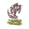
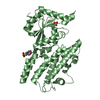
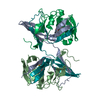
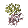
T-cell-specificguaninenucleotidetriphosphate-bindingprotein2 / Interferon-gamma-inducible GTPase Ifggb6 protein / T-cell-specific guanine nucleotide triphosphate- ...Interferon-gamma-inducible GTPase Ifggb6 protein / T-cell-specific guanine nucleotide triphosphate-binding protein
Mass: 47330.719 Da / Num. of mol.: 1 Source method: isolated from a genetically manipulated source Source: (gene. exp.) Mus musculus (house mouse) / Gene: Tgtp2, Ifggb6, Irgb6, Mg21, Tgtp / Production host: Escherichia coli (E. coli) References: UniProt: Q3T9E4, Hydrolases; Acting on acid anhydrides; Acting on GTP to facilitate cellular and subcellular movement
Resolution: 1.51→43.29 Å / Cor.coef. Fo:Fc: 0.978 / Cor.coef. Fo:Fc free: 0.961 / SU B: 3.194 / SU ML: 0.051 / Cross valid method: THROUGHOUT / ESU R: 0.074 / ESU R Free: 0.071 / Stereochemistry target values: MAXIMUM LIKELIHOOD / Details: HYDROGENS HAVE BEEN ADDED IN THE RIDING POSITIONS
Rfactor
Num. reflection
% reflection
Selection details
Rfree
0.19687
1997
2.9 %
RANDOM
Rwork
0.14686
-
-
-
obs
0.14831
65855
99.94 %
-
Solvent computation
Ion probe radii: 0.8 Å / Shrinkage radii: 0.8 Å / VDW probe radii: 1.2 Å / Solvent model: MASK
 Movie
Movie Controller


 Open data
 Basic information
 Components
 Keywords
 Function and homology information
 X-RAY DIFFRACTION /
  Authors
 Japan, 12items
  Citation
 Structure visualization
 Downloads & links
 Other downloads
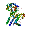


 PDBj
PDBj Assembly


 Mass: 523.180 Da / Num. of mol.: 1 / Source method: obtained synthetically / Formula: C10H16N5O14P3 / Feature type: SUBJECT OF INVESTIGATION / Comment: GTP, energy-carrying molecule*YM
 Mass: 18.015 Da / Num. of mol.: 303 / Source method: isolated from a natural source / Formula: H2O
 Sample preparation
 Processing