 Movie
Movie Controller

[English] 日本語
 Yorodumi
- PDB-7va2: PaOrn Oligoribonuclease D11A mutant with product GMP complex structure -
+ Open data

- Basic information
| Entry | Database: PDB / ID: 7va2 | |||||||||
|---|---|---|---|---|---|---|---|---|---|---|
| Title | PaOrn Oligoribonuclease D11A mutant with product GMP complex structure | |||||||||
 Components Components | Oligoribonuclease | |||||||||
 Keywords Keywords | HYDROLASE / PaOrn / Oligoribonuclease / Pseudomonas aeruginosa. | |||||||||
| Function / homology |  Function and homology information Function and homology informationHydrolases; Acting on ester bonds; Exonucleases that are active with either ribo- or deoxyribonucleic acids and produce 5'-phosphomonoesters / DNA metabolic process / 3'-5'-RNA exonuclease activity / nucleic acid binding / cytoplasm Similarity search - Function | |||||||||
| Biological species |   Pseudomonas aeruginosa (bacteria) Pseudomonas aeruginosa (bacteria) | |||||||||
| Method |  X-RAY DIFFRACTION / SYNCHROTRON / MOLECULAR REPLACEMENT / Resolution: 2.3 Å X-RAY DIFFRACTION / SYNCHROTRON / MOLECULAR REPLACEMENT / Resolution: 2.3 Å | |||||||||
 Authors Authors | Zhang, J. / Zhang, Q. / Bartlam, M. | |||||||||
| Funding support |  China, 2items China, 2items
| |||||||||
 Citation Citation | Journal: To Be Published Title: PaOrn Oligoribonuclease D11A mutant with product GMP complex structure Authors: Zhang, J. / Zhang, Q. / Bartlam, M. | |||||||||
| History |
|
- Structure visualization
| Structure viewer | Molecule: MolmilJmol/JSmol |
|---|
- Downloads & links
-Download
| PDBx/mmCIF format | 7va2.cif.gz | 195.6 KB | Display | PDBx/mmCIF format |
|---|---|---|---|---|
| PDB format | pdb7va2.ent.gz | 129.3 KB | Display | PDB format |
| PDBx/mmJSON format | 7va2.json.gz | Tree view | PDBx/mmJSON format | |
| Others |  Other downloads Other downloads |
-Validation report
| Summary document | 7va2_validation.pdf.gz | 1.4 MB | Display | wwPDB validaton report |
|---|---|---|---|---|
| Full document | 7va2_full_validation.pdf.gz | 1.4 MB | Display | |
| Data in XML | 7va2_validation.xml.gz | 16.2 KB | Display | |
| Data in CIF | 7va2_validation.cif.gz | 21.9 KB | Display | |
| Arichive directory | https://data.pdbj.org/pub/pdb/validation_reports/va/7va2ftp://data.pdbj.org/pub/pdb/validation_reports/va/7va2 | HTTPS FTP |
-Related structure data
| Related structure data | 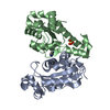 7v9zS S: Starting model for refinement |
|---|---|
| Similar structure data |
-Links
 PDBj
PDBj


- Assembly
| Deposited unit | 
| ||||||||||||
|---|---|---|---|---|---|---|---|---|---|---|---|---|---|
| 1 |
| ||||||||||||
| Unit cell |
|
-Components
| #1: Protein | Mass: 20809.586 Da / Num. of mol.: 2 / Mutation: D11A Source method: isolated from a genetically manipulated source Source: (gene. exp.) Pseudomonas aeruginosa (strain ATCC 15692 / DSM 22644 / CIP 104116 / JCM 14847 / LMG 12228 / 1C / PRS 101 / PAO1) (bacteria)Gene: orn, PA4951 / Production host: References: UniProt: P57665, Hydrolases; Acting on ester bonds #2: Chemical | ChemComp-IMD / |   Mass: 69.085 Da / Num. of mol.: 1 / Source method: obtained synthetically / Formula: C3H5N2 / Feature type: SUBJECT OF INVESTIGATION Mass: 69.085 Da / Num. of mol.: 1 / Source method: obtained synthetically / Formula: C3H5N2 / Feature type: SUBJECT OF INVESTIGATION#3: Chemical | ChemComp-PO4 / |   Mass: 94.971 Da / Num. of mol.: 1 / Source method: obtained synthetically / Formula: PO4 / Feature type: SUBJECT OF INVESTIGATION Mass: 94.971 Da / Num. of mol.: 1 / Source method: obtained synthetically / Formula: PO4 / Feature type: SUBJECT OF INVESTIGATION#4: Chemical | ChemComp-5GP / |   Mass: 363.221 Da / Num. of mol.: 1 / Source method: obtained synthetically / Formula: C10H14N5O8P / Feature type: SUBJECT OF INVESTIGATION Mass: 363.221 Da / Num. of mol.: 1 / Source method: obtained synthetically / Formula: C10H14N5O8P / Feature type: SUBJECT OF INVESTIGATION#5: Water | ChemComp-HOH / |  Mass: 18.015 Da / Num. of mol.: 83 / Source method: isolated from a natural source / Formula: H2O Mass: 18.015 Da / Num. of mol.: 83 / Source method: isolated from a natural source / Formula: H2OHas ligand of interest | Y | Has protein modification | Y | |
|---|
-Experimental details
-Experiment
| Experiment | Method: X-RAY DIFFRACTION / Number of used crystals: 1 |
|---|
- Sample preparation
| Crystal | Density Matthews: 3.94 Å3/Da / Density % sol: 68.79 % |
|---|---|
| Crystal grow | Temperature: 293 K / Method: vapor diffusion / pH: 8 / Details: 1.2M (NH4)2HPO4, 0.1M imidazole pH 8.0 |
-Data collection
| Diffraction | Mean temperature: 100 K / Serial crystal experiment: N |
|---|---|
| Diffraction source | Source: SYNCHROTRON / Site: SSRF / Beamline: BL18U1 / Wavelength: 0.97915 Å |
| Detector | Type: DECTRIS PILATUS3 6M / Detector: PIXEL / Date: Nov 11, 2018 |
| Radiation | Protocol: SINGLE WAVELENGTH / Monochromatic (M) / Laue (L): M / Scattering type: x-ray |
| Radiation wavelength | Wavelength: 0.97915 Å / Relative weight: 1 |
| Reflection | Resolution: 2.3→50 Å / Num. obs: 30350 / % possible obs: 99.8 % / Redundancy: 4.8 % / Biso Wilson estimate: 49.05 Å2 / CC1/2: 0.969 / CC star: 0.992 / Rpim(I) all: 0.092 / Net I/σ(I): 15.61 |
| Reflection shell | Resolution: 2.3→2.34 Å / Redundancy: 4.4 % / Mean I/σ(I) obs: 1.15 / Num. unique obs: 1541 / CC1/2: 0.701 / CC star: 0.908 / % possible all: 100 |
- Processing
| Software |
| ||||||||||||||||||||||||||||||||||||||||||||||||||||||||||||||||||||||||||||||||||||||||||||||||||
|---|---|---|---|---|---|---|---|---|---|---|---|---|---|---|---|---|---|---|---|---|---|---|---|---|---|---|---|---|---|---|---|---|---|---|---|---|---|---|---|---|---|---|---|---|---|---|---|---|---|---|---|---|---|---|---|---|---|---|---|---|---|---|---|---|---|---|---|---|---|---|---|---|---|---|---|---|---|---|---|---|---|---|---|---|---|---|---|---|---|---|---|---|---|---|---|---|---|---|---|
| Refinement | Method to determine structure: MOLECULAR REPLACEMENT Starting model: 7V9Z Resolution: 2.3→34.91 Å / SU ML: 0.2474 / Cross valid method: FREE R-VALUE / σ(F): 1.33 / Phase error: 27.7035 Stereochemistry target values: GeoStd + Monomer Library + CDL v1.2
| ||||||||||||||||||||||||||||||||||||||||||||||||||||||||||||||||||||||||||||||||||||||||||||||||||
| Solvent computation | Shrinkage radii: 0.9 Å / VDW probe radii: 1.11 Å / Solvent model: FLAT BULK SOLVENT MODEL | ||||||||||||||||||||||||||||||||||||||||||||||||||||||||||||||||||||||||||||||||||||||||||||||||||
| Displacement parameters | Biso mean: 63.8 Å2 | ||||||||||||||||||||||||||||||||||||||||||||||||||||||||||||||||||||||||||||||||||||||||||||||||||
| Refinement step | Cycle: LAST / Resolution: 2.3→34.91 Å
| ||||||||||||||||||||||||||||||||||||||||||||||||||||||||||||||||||||||||||||||||||||||||||||||||||
| Refine LS restraints |
| ||||||||||||||||||||||||||||||||||||||||||||||||||||||||||||||||||||||||||||||||||||||||||||||||||
| LS refinement shell |
| ||||||||||||||||||||||||||||||||||||||||||||||||||||||||||||||||||||||||||||||||||||||||||||||||||
| Refinement TLS params. | Method: refined / Origin x: 97.8144293403 Å / Origin y: 89.5912685673 Å / Origin z: 0.910304144951 Å
| ||||||||||||||||||||||||||||||||||||||||||||||||||||||||||||||||||||||||||||||||||||||||||||||||||
| Refinement TLS group | Selection details: all |
