 Movie
Movie Controller

[English] 日本語
 Yorodumi
- PDB-7va1: Crystal structure of human 3-phosphoglycerate dehydrogenase in co... -
+ Open data

- Basic information
| Entry | Database: PDB / ID: 7va1 | ||||||
|---|---|---|---|---|---|---|---|
| Title | Crystal structure of human 3-phosphoglycerate dehydrogenase in complex with GDD-04-35 | ||||||
 Components Components | D-3-phosphoglycerate dehydrogenase | ||||||
 Keywords Keywords | OXIDOREDUCTASE | ||||||
| Function / homology |  Function and homology information Function and homology information2-oxoglutarate reductase / threonine metabolic process / glial cell development / taurine metabolic process / phosphoglycerate dehydrogenase / phosphoglycerate dehydrogenase activity / gamma-aminobutyric acid metabolic process / Serine metabolism / glycine metabolic process / L-serine biosynthetic process ...2-oxoglutarate reductase / threonine metabolic process / glial cell development / taurine metabolic process / phosphoglycerate dehydrogenase / phosphoglycerate dehydrogenase activity / gamma-aminobutyric acid metabolic process / Serine metabolism / glycine metabolic process / L-serine biosynthetic process / malate dehydrogenase / L-malate dehydrogenase (NAD+) activity / glutamine metabolic process / G1 to G0 transition / neural tube development / spinal cord development / brain development / NAD binding / neuron projection development / regulation of gene expression / electron transfer activity / extracellular exosome / cytosol Similarity search - Function | ||||||
| Biological species |  Homo sapiens (human) Homo sapiens (human) | ||||||
| Method |  X-RAY DIFFRACTION / SYNCHROTRON / MOLECULAR REPLACEMENT / Resolution: 1.74 Å X-RAY DIFFRACTION / SYNCHROTRON / MOLECULAR REPLACEMENT / Resolution: 1.74 Å | ||||||
 Authors Authors | Cen, Y. / Gao, D. / Zhou, J. / Tian, P. | ||||||
| Funding support |  China, 1items China, 1items
| ||||||
 Citation Citation | Journal: J.Med.Chem. / Year: 2023 Title: Discovery of Novel Drug-like PHGDH Inhibitors to Disrupt Serine Biosynthesis for Cancer Therapy. Authors: Gao, D. / Tang, S. / Cen, Y. / Yuan, L. / Lan, X. / Li, Q.H. / Lin, G.Q. / Huang, M. / Tian, P. | ||||||
| History |
|
- Structure visualization
| Structure viewer | Molecule: MolmilJmol/JSmol |
|---|
- Downloads & links
-Download
| PDBx/mmCIF format | 7va1.cif.gz | 377.3 KB | Display | PDBx/mmCIF format |
|---|---|---|---|---|
| PDB format | pdb7va1.ent.gz | 255.8 KB | Display | PDB format |
| PDBx/mmJSON format | 7va1.json.gz | Tree view | PDBx/mmJSON format | |
| Others |  Other downloads Other downloads |
-Validation report
| Arichive directory | https://data.pdbj.org/pub/pdb/validation_reports/va/7va1ftp://data.pdbj.org/pub/pdb/validation_reports/va/7va1 | HTTPS FTP |
|---|
-Related structure data
| Related structure data | 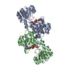 2g76S S: Starting model for refinement |
|---|---|
| Similar structure data |
-Links
 PDBj
PDBj
- Assembly
| Deposited unit | 
| |||||||||||||||||||||||||||||||||||||||||||||||||||||||||||||||||||||||||||||||||||||||||||
|---|---|---|---|---|---|---|---|---|---|---|---|---|---|---|---|---|---|---|---|---|---|---|---|---|---|---|---|---|---|---|---|---|---|---|---|---|---|---|---|---|---|---|---|---|---|---|---|---|---|---|---|---|---|---|---|---|---|---|---|---|---|---|---|---|---|---|---|---|---|---|---|---|---|---|---|---|---|---|---|---|---|---|---|---|---|---|---|---|---|---|---|---|
| 1 | 
| |||||||||||||||||||||||||||||||||||||||||||||||||||||||||||||||||||||||||||||||||||||||||||
| 2 | 
| |||||||||||||||||||||||||||||||||||||||||||||||||||||||||||||||||||||||||||||||||||||||||||
| 3 | 
| |||||||||||||||||||||||||||||||||||||||||||||||||||||||||||||||||||||||||||||||||||||||||||
| 4 | 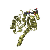
| |||||||||||||||||||||||||||||||||||||||||||||||||||||||||||||||||||||||||||||||||||||||||||
| Unit cell |
| |||||||||||||||||||||||||||||||||||||||||||||||||||||||||||||||||||||||||||||||||||||||||||
| Noncrystallographic symmetry (NCS) | NCS domain:
NCS domain segments: Ens-ID: 1
|