 Movie
Movie Controller


+ Open data

- Basic information
| Entry | Database: PDB / ID: 7v5s | ||||||
|---|---|---|---|---|---|---|---|
| Title | Crystal structure of human bleomycin hydrolase C73A mutant | ||||||
 Components Components | Bleomycin hydrolase | ||||||
 Keywords Keywords | HYDROLASE / cysteine protease | ||||||
| Function / homology |  Function and homology information Function and homology informationbleomycin hydrolase / cysteine-type aminopeptidase activity / L-homocysteine catabolic process / aminopeptidase activity / carboxypeptidase activity / cysteine-type peptidase activity / response to toxic substance / protein polyubiquitination / Antigen processing: Ubiquitination & Proteasome degradation / response to xenobiotic stimulus ...bleomycin hydrolase / cysteine-type aminopeptidase activity / L-homocysteine catabolic process / aminopeptidase activity / carboxypeptidase activity / cysteine-type peptidase activity / response to toxic substance / protein polyubiquitination / Antigen processing: Ubiquitination & Proteasome degradation / response to xenobiotic stimulus / cysteine-type endopeptidase activity / proteolysis / extracellular exosome / identical protein binding / nucleus / cytoplasm / cytosol Similarity search - Function | ||||||
| Biological species |  Homo sapiens (human) Homo sapiens (human) | ||||||
| Method |  X-RAY DIFFRACTION / SYNCHROTRON / MOLECULAR REPLACEMENT / Resolution: 3.02 Å X-RAY DIFFRACTION / SYNCHROTRON / MOLECULAR REPLACEMENT / Resolution: 3.02 Å | ||||||
 Authors Authors | Chang, C.Y. / Zheng, Y.Z. / Huang, S.J. / Wang, Y.L. / Toh, S.I. / Lin, E.C. | ||||||
| Funding support |  Taiwan, 1items Taiwan, 1items
| ||||||
 Citation Citation | Journal: Chembiochem / Year: 2022 Title: The Structure-Function Relationship of Human Bleomycin Hydrolase: Mutation of a Cysteine Protease into a Serine Protease. Authors: Zheng, Y.Z. / Cui, J. / Wang, Y.L. / Huang, S.J. / Lin, E.C. / Huang, S.C. / Rudolf, J.D. / Yan, X. / Chang, C.Y. | ||||||
| History |
|
- Structure visualization
| Structure viewer | Molecule: MolmilJmol/JSmol |
|---|
- Downloads & links
-Download
| PDBx/mmCIF format | 7v5s.cif.gz | 108.4 KB | Display | PDBx/mmCIF format |
|---|---|---|---|---|
| PDB format | pdb7v5s.ent.gz | 80.9 KB | Display | PDB format |
| PDBx/mmJSON format | 7v5s.json.gz | Tree view | PDBx/mmJSON format | |
| Others |  Other downloads Other downloads |
-Validation report
| Arichive directory | https://data.pdbj.org/pub/pdb/validation_reports/v5/7v5sftp://data.pdbj.org/pub/pdb/validation_reports/v5/7v5s | HTTPS FTP |
|---|
-Related structure data
| Related structure data |  7v5lSC 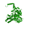 7v5tC 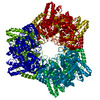 7xf9C S: Starting model for refinement C: citing same article ( |
|---|---|
| Similar structure data |
-Links
 PDBj
PDBj
- Assembly
| Deposited unit | 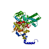
| ||||||||
|---|---|---|---|---|---|---|---|---|---|
| 1 | x 6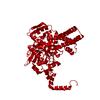
| ||||||||
| Unit cell |
|
-Components
| #1: Protein | Mass: 54767.387 Da / Num. of mol.: 1 / Mutation: C73A Source method: isolated from a genetically manipulated source Source: (gene. exp.) Homo sapiens (human) / Gene: BLMH / Production host:  |
|---|---|
| #2: Chemical | ChemComp-PG4 /   Mass: 194.226 Da / Num. of mol.: 1 / Source method: obtained synthetically / Formula: C8H18O5 / Feature type: SUBJECT OF INVESTIGATION / Comment: precipitant*YM Mass: 194.226 Da / Num. of mol.: 1 / Source method: obtained synthetically / Formula: C8H18O5 / Feature type: SUBJECT OF INVESTIGATION / Comment: precipitant*YM |
| #3: Chemical | ChemComp-PGE /   Mass: 150.173 Da / Num. of mol.: 1 / Source method: obtained synthetically / Formula: C6H14O4 / Feature type: SUBJECT OF INVESTIGATION Mass: 150.173 Da / Num. of mol.: 1 / Source method: obtained synthetically / Formula: C6H14O4 / Feature type: SUBJECT OF INVESTIGATION |
| #4: Water | ChemComp-HOH /  Mass: 18.015 Da / Num. of mol.: 19 / Source method: isolated from a natural source / Formula: H2O Mass: 18.015 Da / Num. of mol.: 19 / Source method: isolated from a natural source / Formula: H2O |
| Has ligand of interest | Y |
-Experimental details
-Experiment
| Experiment | Method: X-RAY DIFFRACTION / Number of used crystals: 1 |
|---|
- Sample preparation
| Crystal | Density Matthews: 12.26 Å3/Da / Density % sol: 89.97 % |
|---|---|
| Crystal grow | Temperature: 293 K / Method: vapor diffusion, hanging drop / Details: 30% (w/v) PEG 400, 0.1mM CHES, pH 9.5 |
-Data collection
| Diffraction | Mean temperature: 100 K / Serial crystal experiment: N |
|---|---|
| Diffraction source | Source: SYNCHROTRON / Site: NSRRC / Beamline: BL13B1 / Wavelength: 0.9732 Å |
| Detector | Type: ADSC QUANTUM 315r / Detector: CCD / Date: Apr 8, 2021 |
| Radiation | Protocol: SINGLE WAVELENGTH / Monochromatic (M) / Laue (L): M / Scattering type: x-ray |
| Radiation wavelength | Wavelength: 0.9732 Å / Relative weight: 1 |
| Reflection | Resolution: 3.02→30 Å / Num. obs: 53491 / % possible obs: 100 % / Redundancy: 11.3 % / CC1/2: 0.98 / Net I/σ(I): 14.2 |
| Reflection shell | Resolution: 3.02→3.13 Å / Redundancy: 11.9 % / Mean I/σ(I) obs: 2.7 / Num. unique obs: 5272 / CC1/2: 0.865 / % possible all: 100 |
- Processing
| Software |
| ||||||||||||||||||||||||||||||||||||||||||||||||||||||||||||
|---|---|---|---|---|---|---|---|---|---|---|---|---|---|---|---|---|---|---|---|---|---|---|---|---|---|---|---|---|---|---|---|---|---|---|---|---|---|---|---|---|---|---|---|---|---|---|---|---|---|---|---|---|---|---|---|---|---|---|---|---|---|
| Refinement | Method to determine structure: MOLECULAR REPLACEMENT Starting model: 7V5L Resolution: 3.02→29.83 Å / Cor.coef. Fo:Fc: 0.951 / Cor.coef. Fo:Fc free: 0.938 / SU B: 7.095 / SU ML: 0.118 / Cross valid method: THROUGHOUT / σ(F): 0 / ESU R: 0.179 / ESU R Free: 0.164 / Stereochemistry target values: MAXIMUM LIKELIHOOD Details: HYDROGENS HAVE BEEN ADDED IN THE RIDING POSITIONS U VALUES : REFINED INDIVIDUALLY
| ||||||||||||||||||||||||||||||||||||||||||||||||||||||||||||
| Solvent computation | Ion probe radii: 0.8 Å / Shrinkage radii: 0.8 Å / VDW probe radii: 1.2 Å / Solvent model: MASK | ||||||||||||||||||||||||||||||||||||||||||||||||||||||||||||
| Displacement parameters | Biso max: 144.08 Å2 / Biso mean: 63.93 Å2 / Biso min: 39.38 Å2
| ||||||||||||||||||||||||||||||||||||||||||||||||||||||||||||
| Refinement step | Cycle: final / Resolution: 3.02→29.83 Å
| ||||||||||||||||||||||||||||||||||||||||||||||||||||||||||||
| Refine LS restraints |
| ||||||||||||||||||||||||||||||||||||||||||||||||||||||||||||
| LS refinement shell | Resolution: 3.023→3.101 Å / Rfactor Rfree error: 0 / Total num. of bins used: 20
|
