 Movie
Movie Controller

[English] 日本語
 Yorodumi
- PDB-7v55: Crystal structure of phospholipase D from Pseudomonas aeruginosa ... -
+ Open data

- Basic information
| Entry | Database: PDB / ID: 7v55 | ||||||
|---|---|---|---|---|---|---|---|
| Title | Crystal structure of phospholipase D from Pseudomonas aeruginosa PAO1 using in situ proteolysis | ||||||
 Components Components | Phospholipase D | ||||||
 Keywords Keywords | HYDROLASE / phospholipase D / Toxin | ||||||
| Function / homology | Phospholipase D family / Phospholipase D Active site motif / Phospholipase D. Active site motifs. / Phospholipase D/Transphosphatidylase / Phospholipase D phosphodiesterase active site profile. / phospholipid catabolic process / phospholipase D activity / metal ion binding / Phospholipase D Function and homology information Function and homology information | ||||||
| Biological species |   Pseudomonas aeruginosa (bacteria) Pseudomonas aeruginosa (bacteria) | ||||||
| Method |  X-RAY DIFFRACTION / SYNCHROTRON / MOLECULAR REPLACEMENT / Resolution: 3 Å X-RAY DIFFRACTION / SYNCHROTRON / MOLECULAR REPLACEMENT / Resolution: 3 Å | ||||||
 Authors Authors | Yang, Y. / Li, Z. | ||||||
| Funding support | 1items
| ||||||
 Citation Citation | Journal: Nat Commun / Year: 2022 Title: Structural insights into PA3488-mediated inactivation of Pseudomonas aeruginosa PldA. Authors: Xiaoyun Yang / Zongqiang Li / Liang Zhao / Zhun She / Zengqiang Gao / Sen-Fang Sui / Yuhui Dong / Yanhua Li /  Abstract: PldA, a phospholipase D (PLD) effector, catalyzes hydrolysis of the phosphodiester bonds of glycerophospholipids-the main component of cell membranes-and assists the invasion of the opportunistic ...PldA, a phospholipase D (PLD) effector, catalyzes hydrolysis of the phosphodiester bonds of glycerophospholipids-the main component of cell membranes-and assists the invasion of the opportunistic pathogen Pseudomonas aeruginosa. As a cognate immunity protein, PA3488 can inhibit the activity of PldA to avoid self-toxicity. However, the precise inhibitory mechanism remains elusive. We determine the crystal structures of full-length and truncated PldA and the cryogenic electron microscopy structure of the PldA-PA3488 complex. Structural analysis reveals that there are different intermediates of PldA between the "open" and "closed" states of the catalytic pocket, accompanied by significant conformational changes in the "lid" region and the peripheral helical domain. Through structure-based mutational analysis, we identify the key residues responsible for the enzymatic activity of PldA. Together, these data provide an insight into the molecular mechanisms of PldA invasion and its neutralization by PA3488, aiding future design of PLD-targeted inhibitors and drugs. | ||||||
| History |
|
- Structure visualization
| Structure viewer | Molecule: MolmilJmol/JSmol |
|---|
- Downloads & links
-Download
| PDBx/mmCIF format | 7v55.cif.gz | 171.5 KB | Display | PDBx/mmCIF format |
|---|---|---|---|---|
| PDB format | pdb7v55.ent.gz | 128.4 KB | Display | PDB format |
| PDBx/mmJSON format | 7v55.json.gz | Tree view | PDBx/mmJSON format | |
| Others |  Other downloads Other downloads |
-Validation report
| Summary document | 7v55_validation.pdf.gz | 435.4 KB | Display | wwPDB validaton report |
|---|---|---|---|---|
| Full document | 7v55_full_validation.pdf.gz | 444.5 KB | Display | |
| Data in XML | 7v55_validation.xml.gz | 28.1 KB | Display | |
| Data in CIF | 7v55_validation.cif.gz | 37.9 KB | Display | |
| Arichive directory | https://data.pdbj.org/pub/pdb/validation_reports/v5/7v55ftp://data.pdbj.org/pub/pdb/validation_reports/v5/7v55 | HTTPS FTP |
-Related structure data
| Related structure data |  7v53SC  7wdkC S: Starting model for refinement C: citing same article ( |
|---|---|
| Similar structure data |
-Links
 PDBj
PDBj- Assembly
| Deposited unit | 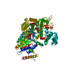
| ||||||||
|---|---|---|---|---|---|---|---|---|---|
| 1 |
| ||||||||
| Unit cell |
|
-Components
| #1: Protein | Mass: 122478.062 Da / Num. of mol.: 1 Source method: isolated from a genetically manipulated source Source: (gene. exp.) Pseudomonas aeruginosa (strain ATCC 15692 / DSM 22644 / CIP 104116 / JCM 14847 / LMG 12228 / 1C / PRS 101 / PAO1) (bacteria)Gene: pldA, PA3487 / Production host: |
|---|---|
| #2: Chemical | ChemComp-CA /   Mass: 40.078 Da / Num. of mol.: 1 / Source method: obtained synthetically / Formula: Ca Mass: 40.078 Da / Num. of mol.: 1 / Source method: obtained synthetically / Formula: Ca |
| #3: Water | ChemComp-HOH /  Mass: 18.015 Da / Num. of mol.: 3 / Source method: isolated from a natural source / Formula: H2O Mass: 18.015 Da / Num. of mol.: 3 / Source method: isolated from a natural source / Formula: H2O |
| Has ligand of interest | N |
-Experimental details
-Experiment
| Experiment | Method: X-RAY DIFFRACTION / Number of used crystals: 1 |
|---|
- Sample preparation
| Crystal | Density Matthews: 1.99 Å3/Da / Density % sol: 38.33 % |
|---|---|
| Crystal grow | Temperature: 291.1 K / Method: evaporation Details: 0.2M Potassium phosphate dibasic, 20% w/v Polyethylene glycol 3350 |
-Data collection
| Diffraction | Mean temperature: 100 K / Serial crystal experiment: N | ||||||||||||||||||||||||||||||
|---|---|---|---|---|---|---|---|---|---|---|---|---|---|---|---|---|---|---|---|---|---|---|---|---|---|---|---|---|---|---|---|
| Diffraction source | Source: SYNCHROTRON / Site: SSRF / Beamline: BL17U1 / Wavelength: 0.97853 Å | ||||||||||||||||||||||||||||||
| Detector | Type: ADSC QUANTUM 315r / Detector: CCD / Date: Apr 26, 2019 | ||||||||||||||||||||||||||||||
| Radiation | Protocol: SINGLE WAVELENGTH / Monochromatic (M) / Laue (L): M / Scattering type: x-ray | ||||||||||||||||||||||||||||||
| Radiation wavelength | Wavelength: 0.97853 Å / Relative weight: 1 | ||||||||||||||||||||||||||||||
| Reflection | Resolution: 3→72.37 Å / Num. obs: 19242 / % possible obs: 94.4 % / Redundancy: 5.3 % / Biso Wilson estimate: 68.27 Å2 / CC1/2: 0.996 / Rmerge(I) obs: 0.123 / Rpim(I) all: 0.057 / Rrim(I) all: 0.136 / Net I/σ(I): 11.8 / Num. measured all: 102295 / Scaling rejects: 85 | ||||||||||||||||||||||||||||||
| Reflection shell | Diffraction-ID: 1
|
- Processing
| Software |
| ||||||||||||||||||||||||||||||||||||||||||||||||||||||||
|---|---|---|---|---|---|---|---|---|---|---|---|---|---|---|---|---|---|---|---|---|---|---|---|---|---|---|---|---|---|---|---|---|---|---|---|---|---|---|---|---|---|---|---|---|---|---|---|---|---|---|---|---|---|---|---|---|---|
| Refinement | Method to determine structure: MOLECULAR REPLACEMENT Starting model: 7V53 Resolution: 3→69.34 Å / SU ML: 0.35 / Cross valid method: THROUGHOUT / σ(F): 1.34 / Phase error: 24.47 / Stereochemistry target values: ML
| ||||||||||||||||||||||||||||||||||||||||||||||||||||||||
| Solvent computation | Shrinkage radii: 0.9 Å / VDW probe radii: 1.11 Å / Solvent model: FLAT BULK SOLVENT MODEL | ||||||||||||||||||||||||||||||||||||||||||||||||||||||||
| Displacement parameters | Biso max: 152.93 Å2 / Biso mean: 60.7261 Å2 / Biso min: 25.26 Å2 | ||||||||||||||||||||||||||||||||||||||||||||||||||||||||
| Refinement step | Cycle: final / Resolution: 3→69.34 Å
| ||||||||||||||||||||||||||||||||||||||||||||||||||||||||
| LS refinement shell | Refine-ID: X-RAY DIFFRACTION / Rfactor Rfree error: 0 / Total num. of bins used: 7
|
