 Movie
Movie Controller

[English] 日本語
 Yorodumi
- PDB-7v50: Structure of cyclohexanone monooxygenase mutant from Acinetobacte... -
+ Open data

- Basic information
| Entry | Database: PDB / ID: 7v50 | |||||||||
|---|---|---|---|---|---|---|---|---|---|---|
| Title | Structure of cyclohexanone monooxygenase mutant from Acinetobacter calcoaceticus | |||||||||
 Components Components | Putative flavin-binding monooxygenase | |||||||||
 Keywords Keywords | OXIDOREDUCTASE / BVMO_positive mutant | |||||||||
| Function / homology |  Function and homology information Function and homology informationN,N-dimethylaniline monooxygenase activity / NADP binding / flavin adenine dinucleotide binding Similarity search - Function | |||||||||
| Biological species |  Acinetobacter calcoaceticus (bacteria) Acinetobacter calcoaceticus (bacteria) | |||||||||
| Method |  X-RAY DIFFRACTION / SYNCHROTRON / MOLECULAR REPLACEMENT / Resolution: 2.3 Å X-RAY DIFFRACTION / SYNCHROTRON / MOLECULAR REPLACEMENT / Resolution: 2.3 Å | |||||||||
 Authors Authors | Wu, Y. / Yu, H. | |||||||||
| Funding support | 1items
| |||||||||
 Citation Citation | Journal: CHINESE J CATAL / Year: 2023 Title: Precise regulation of the substrate selectivity of Baeyer-Villiger monooxygenase to minimize overoxidation of prazole sulfoxides. Authors: Wu, Y. / Chen, Q.Q. / Chen, Q. / Geng, Q. / Zhang, Q. / Zheng, Y.C. / Zhao, C. / Zhang, Y. / Zhou, J. / Wang, B. / Xu, J.H. / Yu, H.L. | |||||||||
| History |
|
- Structure visualization
| Structure viewer | Molecule: MolmilJmol/JSmol |
|---|
- Downloads & links
-Download
| PDBx/mmCIF format | 7v50.cif.gz | 129.8 KB | Display | PDBx/mmCIF format |
|---|---|---|---|---|
| PDB format | pdb7v50.ent.gz | 94.9 KB | Display | PDB format |
| PDBx/mmJSON format | 7v50.json.gz | Tree view | PDBx/mmJSON format | |
| Others |  Other downloads Other downloads |
-Validation report
| Arichive directory | https://data.pdbj.org/pub/pdb/validation_reports/v5/7v50ftp://data.pdbj.org/pub/pdb/validation_reports/v5/7v50 | HTTPS FTP |
|---|
-Related structure data
| Related structure data | 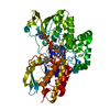 7v4xC  7v51C  6a37S S: Starting model for refinement C: citing same article ( |
|---|---|
| Similar structure data |
-Links
 PDBj
PDBj- Assembly
| Deposited unit | 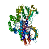
| ||||||||
|---|---|---|---|---|---|---|---|---|---|
| 1 |
| ||||||||
| Unit cell |
|
-Components
| #1: Protein | Mass: 63330.574 Da / Num. of mol.: 1 Mutation: F246Y,F277L,K326C,N386S,I388K,M390I,L426F,F432L,T433A,L435S,S438I,E488K,S489C,W490R,F505L Source method: isolated from a genetically manipulated source Source: (gene. exp.) Acinetobacter calcoaceticus (bacteria) / Gene: P23_1101Production host: References: UniProt: A0A0A8XFY0 |
|---|---|
| #2: Chemical | ChemComp-FAD /   Mass: 785.550 Da / Num. of mol.: 1 / Source method: obtained synthetically / Formula: C27H33N9O15P2 / Comment: FAD*YM Mass: 785.550 Da / Num. of mol.: 1 / Source method: obtained synthetically / Formula: C27H33N9O15P2 / Comment: FAD*YM |
| #3: Chemical | ChemComp-NAP /   Mass: 743.405 Da / Num. of mol.: 1 / Source method: obtained synthetically / Formula: C21H28N7O17P3 Mass: 743.405 Da / Num. of mol.: 1 / Source method: obtained synthetically / Formula: C21H28N7O17P3 |
| #4: Water | ChemComp-HOH /  Mass: 18.015 Da / Num. of mol.: 213 / Source method: isolated from a natural source / Formula: H2O Mass: 18.015 Da / Num. of mol.: 213 / Source method: isolated from a natural source / Formula: H2O |
| Has ligand of interest | N |
-Experimental details
-Experiment
| Experiment | Method: X-RAY DIFFRACTION / Number of used crystals: 1 |
|---|
- Sample preparation
| Crystal | Density Matthews: 2.36 Å3/Da / Density % sol: 47.93 % |
|---|---|
| Crystal grow | Temperature: 289 K / Method: vapor diffusion, sitting drop Details: bicine/Trizma base, PEG20000, PEG MME 550, 1,6-Hexanediol, 1-Butanal, 1,2-Propanediol, 1,4-Butanediol, 1,3-Propanediol |
-Data collection
| Diffraction | Mean temperature: 80 K / Serial crystal experiment: N |
|---|---|
| Diffraction source | Source: SYNCHROTRON / Site: SSRF  / Beamline: BL19U1 / Wavelength: 0.987 Å / Beamline: BL19U1 / Wavelength: 0.987 Å |
| Detector | Type: ADSC QUANTUM 315r / Detector: CCD / Date: Oct 1, 2018 |
| Radiation | Protocol: SINGLE WAVELENGTH / Monochromatic (M) / Laue (L): M / Scattering type: x-ray |
| Radiation wavelength | Wavelength: 0.987 Å / Relative weight: 1 |
| Reflection | Resolution: 2.07→50 Å / Num. obs: 34734 / % possible obs: 99.9 % / Redundancy: 6.1 % / Rmerge(I) obs: 0.07 / Net I/σ(I): 33.75 |
| Reflection shell | Resolution: 2.08→2.12 Å / Rmerge(I) obs: 0.07 / Num. unique obs: 34734 |
- Processing
| Software |
| ||||||||||||||||||||||||||||||||||||||||||||||||||||||||||||||||||||||
|---|---|---|---|---|---|---|---|---|---|---|---|---|---|---|---|---|---|---|---|---|---|---|---|---|---|---|---|---|---|---|---|---|---|---|---|---|---|---|---|---|---|---|---|---|---|---|---|---|---|---|---|---|---|---|---|---|---|---|---|---|---|---|---|---|---|---|---|---|---|---|---|
| Refinement | Method to determine structure: MOLECULAR REPLACEMENT Starting model: 6A37 Resolution: 2.3→38.87 Å / SU ML: 0.32 / Cross valid method: FREE R-VALUE / σ(F): 1.36 / Phase error: 26.7 / Stereochemistry target values: ML
| ||||||||||||||||||||||||||||||||||||||||||||||||||||||||||||||||||||||
| Solvent computation | Shrinkage radii: 0.9 Å / VDW probe radii: 1.11 Å / Solvent model: FLAT BULK SOLVENT MODEL | ||||||||||||||||||||||||||||||||||||||||||||||||||||||||||||||||||||||
| Refinement step | Cycle: LAST / Resolution: 2.3→38.87 Å
| ||||||||||||||||||||||||||||||||||||||||||||||||||||||||||||||||||||||
| Refine LS restraints |
| ||||||||||||||||||||||||||||||||||||||||||||||||||||||||||||||||||||||
| LS refinement shell |
|
