 Movie
Movie Controller

[English] 日本語
 Yorodumi
- PDB-7v3n: The complex structure of soBcmB-D307A and its natural precursor 2 -
+ Open data

- Basic information
| Entry | Database: PDB / ID: 7v3n | ||||||
|---|---|---|---|---|---|---|---|
| Title | The complex structure of soBcmB-D307A and its natural precursor 2 | ||||||
 Components Components | dioxygenase | ||||||
 Keywords Keywords | OXIDOREDUCTASE / complex / mutant / bicyclomycin | ||||||
| Function / homology | Chem-5UQ / 2-OXOGLUTARIC ACID / :  Function and homology information Function and homology information | ||||||
| Biological species |  Streptomyces ossamyceticus (bacteria) Streptomyces ossamyceticus (bacteria) | ||||||
| Method |  X-RAY DIFFRACTION / SYNCHROTRON / MOLECULAR REPLACEMENT / Resolution: 1.85001100152 Å X-RAY DIFFRACTION / SYNCHROTRON / MOLECULAR REPLACEMENT / Resolution: 1.85001100152 Å | ||||||
 Authors Authors | Wu, L. / Zhou, J.H. | ||||||
| Funding support | 1items
| ||||||
 Citation Citation | Journal: Nat Catal / Year: 2023 Title: Enzymatic catalysis favours eight-membered over five-membered ring closure in bicyclomycin biosynthesis Authors: He, J.B. / Wu, L. / Wei, W. / Meng, S. / Liu, Z.T. / Wu, X. / Pan, H.X. / Yang, S. / Liang, Y. / Zhou, J. / Tang, G.L. | ||||||
| History |
|
- Structure visualization
| Structure viewer | Molecule: MolmilJmol/JSmol |
|---|
- Downloads & links
-Download
| PDBx/mmCIF format | 7v3n.cif.gz | 101.4 KB | Display | PDBx/mmCIF format |
|---|---|---|---|---|
| PDB format | pdb7v3n.ent.gz | 61.6 KB | Display | PDB format |
| PDBx/mmJSON format | 7v3n.json.gz | Tree view | PDBx/mmJSON format | |
| Others |  Other downloads Other downloads |
-Validation report
| Arichive directory | https://data.pdbj.org/pub/pdb/validation_reports/v3/7v3nftp://data.pdbj.org/pub/pdb/validation_reports/v3/7v3n | HTTPS FTP |
|---|
-Related structure data
| Related structure data | 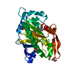 7v2tC  7v2uC  7v2xC 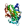 7v34C  7v36C 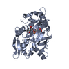 7v3eC  7v3oSC  8hivC S: Starting model for refinement C: citing same article ( |
|---|---|
| Similar structure data |
-Links
 PDBj
PDBj


- Assembly
| Deposited unit | 
| ||||||||||||
|---|---|---|---|---|---|---|---|---|---|---|---|---|---|
| 1 |
| ||||||||||||
| Unit cell |
| ||||||||||||
| Components on special symmetry positions |
|
-Components
-Protein , 1 types, 1 molecules A
| #1: Protein | Mass: 37247.168 Da / Num. of mol.: 1 Source method: isolated from a genetically manipulated source Source: (gene. exp.) Streptomyces ossamyceticus (bacteria) / Production host: |
|---|
-Non-polymers , 5 types, 188 molecules 


| #2: Chemical | ChemComp-FE2 /  Mass: 55.845 Da / Num. of mol.: 1 / Source method: obtained synthetically / Formula: Fe Mass: 55.845 Da / Num. of mol.: 1 / Source method: obtained synthetically / Formula: Fe |
|---|---|
| #3: Chemical | ChemComp-5UQ / ( Mass: 274.313 Da / Num. of mol.: 1 / Source method: obtained synthetically / Formula: C12H22N2O5 / Feature type: SUBJECT OF INVESTIGATION Mass: 274.313 Da / Num. of mol.: 1 / Source method: obtained synthetically / Formula: C12H22N2O5 / Feature type: SUBJECT OF INVESTIGATION |
| #4: Chemical | ChemComp-AKG /  Mass: 146.098 Da / Num. of mol.: 1 / Source method: obtained synthetically / Formula: C5H6O5 / Feature type: SUBJECT OF INVESTIGATION Mass: 146.098 Da / Num. of mol.: 1 / Source method: obtained synthetically / Formula: C5H6O5 / Feature type: SUBJECT OF INVESTIGATION |
| #5: Chemical | ChemComp-GOL /  Mass: 92.094 Da / Num. of mol.: 1 / Source method: obtained synthetically / Formula: C3H8O3 Mass: 92.094 Da / Num. of mol.: 1 / Source method: obtained synthetically / Formula: C3H8O3 |
| #6: Water | ChemComp-HOH / Mass: 18.015 Da / Num. of mol.: 184 / Source method: isolated from a natural source / Formula: H2O |
-Details
| Has ligand of interest | Y |
|---|
-Experimental details
-Experiment
| Experiment | Method: X-RAY DIFFRACTION / Number of used crystals: 1 |
|---|
- Sample preparation
| Crystal | Density Matthews: 2.44 Å3/Da / Density % sol: 49.6 % |
|---|---|
| Crystal grow | Temperature: 293 K / Method: vapor diffusion, sitting drop Details: 0.1M MES monohydrate pH 6.5, 1.6M Ammonium sulfate, 10% 1.4-dioxane |
-Data collection
| Diffraction | Mean temperature: 100 K / Serial crystal experiment: N |
|---|---|
| Diffraction source | Source: SYNCHROTRON / Site: SSRF  / Beamline: BL17U1 / Wavelength: 0.97913 Å / Beamline: BL17U1 / Wavelength: 0.97913 Å |
| Detector | Type: DECTRIS PILATUS3 6M / Detector: PIXEL / Date: Jun 6, 2020 |
| Radiation | Protocol: SINGLE WAVELENGTH / Monochromatic (M) / Laue (L): M / Scattering type: x-ray |
| Radiation wavelength | Wavelength: 0.97913 Å / Relative weight: 1 |
| Reflection | Resolution: 1.85→80.94 Å / Num. obs: 32011 / % possible obs: 99.7 % / Redundancy: 18.8 % / Biso Wilson estimate: 24.8642105118 Å2 / CC1/2: 0.999 / Rmerge(I) obs: 0.09 / Net I/σ(I): 22.1 |
| Reflection shell | Resolution: 1.85→1.9 Å / Rmerge(I) obs: 0.561 / Num. unique obs: 2240 / CC1/2: 0.799 |
- Processing
| Software |
| |||||||||||||||||||||||||||||||||||||||||||||||||||||||||||||||||||||||||||||||||||||||||||
|---|---|---|---|---|---|---|---|---|---|---|---|---|---|---|---|---|---|---|---|---|---|---|---|---|---|---|---|---|---|---|---|---|---|---|---|---|---|---|---|---|---|---|---|---|---|---|---|---|---|---|---|---|---|---|---|---|---|---|---|---|---|---|---|---|---|---|---|---|---|---|---|---|---|---|---|---|---|---|---|---|---|---|---|---|---|---|---|---|---|---|---|---|
| Refinement | Method to determine structure: MOLECULAR REPLACEMENT Starting model: 7V3O Resolution: 1.85001100152→80.9354421746 Å / SU ML: 0.135404379973 / Cross valid method: FREE R-VALUE / σ(F): 1.36071500929 / Phase error: 17.458799539 Stereochemistry target values: GeoStd + Monomer Library + CDL v1.2
| |||||||||||||||||||||||||||||||||||||||||||||||||||||||||||||||||||||||||||||||||||||||||||
| Solvent computation | Shrinkage radii: 0.9 Å / VDW probe radii: 1.11 Å / Solvent model: FLAT BULK SOLVENT MODEL | |||||||||||||||||||||||||||||||||||||||||||||||||||||||||||||||||||||||||||||||||||||||||||
| Displacement parameters | Biso mean: 28.7016062295 Å2 | |||||||||||||||||||||||||||||||||||||||||||||||||||||||||||||||||||||||||||||||||||||||||||
| Refinement step | Cycle: LAST / Resolution: 1.85001100152→80.9354421746 Å
| |||||||||||||||||||||||||||||||||||||||||||||||||||||||||||||||||||||||||||||||||||||||||||
| Refine LS restraints |
| |||||||||||||||||||||||||||||||||||||||||||||||||||||||||||||||||||||||||||||||||||||||||||
| LS refinement shell |
|
