 Movie
Movie Controller

[English] 日本語
 Yorodumi
- PDB-7uyy: The crystal structure of the Pseudomonas aeruginosa aldehyde dehy... -
+ Open data

- Basic information
| Entry | Database: PDB / ID: 7uyy | |||||||||
|---|---|---|---|---|---|---|---|---|---|---|
| Title | The crystal structure of the Pseudomonas aeruginosa aldehyde dehydrogenase encoded by the PA4189 gene in complex with NADH | |||||||||
 Components Components | Aldehyde dehydrogenase | |||||||||
 Keywords Keywords | OXIDOREDUCTASE / tetramer / aldehyde dehydrogenase fold / aminoaldehyde dehydrogenase / ALDH27 family | |||||||||
| Function / homology |  Function and homology information Function and homology informationoxidoreductase activity, acting on the aldehyde or oxo group of donors, NAD or NADP as acceptor / nucleotide binding Similarity search - Function | |||||||||
| Biological species |  Pseudomonas aeruginosa PAO1 (bacteria) Pseudomonas aeruginosa PAO1 (bacteria) | |||||||||
| Method |  X-RAY DIFFRACTION / SYNCHROTRON / MOLECULAR REPLACEMENT / Resolution: 2.7 Å X-RAY DIFFRACTION / SYNCHROTRON / MOLECULAR REPLACEMENT / Resolution: 2.7 Å | |||||||||
 Authors Authors | Gonzalez-Segura, L. / Juarez-Vazquez, A.L. / Munoz-Clares, R.A. | |||||||||
| Funding support |  Mexico, 2items Mexico, 2items
| |||||||||
 Citation Citation | Journal: Biochem.J. / Year: 2023 Title: The uncharacterized Pseudomonas aeruginosa PA4189 is a novel and efficient aminoacetaldehyde dehydrogenase. Authors: Fernandez-Silva, A. / Juarez-Vazquez, A.L. / Gonzalez-Segura, L. / Juarez-Diaz, J.A. / Munoz-Clares, R.A. | |||||||||
| History |
|
- Structure visualization
| Structure viewer | Molecule: MolmilJmol/JSmol |
|---|
- Downloads & links
-Download
| PDBx/mmCIF format | 7uyy.cif.gz | 221.5 KB | Display | PDBx/mmCIF format |
|---|---|---|---|---|
| PDB format | pdb7uyy.ent.gz | 173 KB | Display | PDB format |
| PDBx/mmJSON format | 7uyy.json.gz | Tree view | PDBx/mmJSON format | |
| Others |  Other downloads Other downloads |
-Validation report
| Summary document | 7uyy_validation.pdf.gz | 1.2 MB | Display | wwPDB validaton report |
|---|---|---|---|---|
| Full document | 7uyy_full_validation.pdf.gz | 1.2 MB | Display | |
| Data in XML | 7uyy_validation.xml.gz | 42.7 KB | Display | |
| Data in CIF | 7uyy_validation.cif.gz | 61.6 KB | Display | |
| Arichive directory | https://data.pdbj.org/pub/pdb/validation_reports/uy/7uyyftp://data.pdbj.org/pub/pdb/validation_reports/uy/7uyy | HTTPS FTP |
-Related structure data
| Related structure data | 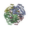 6b4rS S: Starting model for refinement |
|---|---|
| Similar structure data |
-Links
 PDBj
PDBj


- Assembly
| Deposited unit | 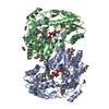
| |||||||||||||||
|---|---|---|---|---|---|---|---|---|---|---|---|---|---|---|---|---|
| 1 | 
| |||||||||||||||
| Unit cell |
| |||||||||||||||
| Components on special symmetry positions |
|
-Components
-Protein , 1 types, 2 molecules AB
| #1: Protein | Mass: 55398.633 Da / Num. of mol.: 2 Source method: isolated from a genetically manipulated source Source: (gene. exp.) Pseudomonas aeruginosa PAO1 (bacteria)Strain: ATCC 15692 / DSM 22644 / CIP 104116 / JCM 14847 / LMG 12228 / 1C / PRS 101 / PAO1 Gene: PA4189 / Plasmid: pET28b-PA4189 / Production host: |
|---|
-Non-polymers , 7 types, 570 molecules 


| #2: Chemical | ChemComp-K /  Mass: 39.098 Da / Num. of mol.: 6 / Source method: obtained synthetically / Formula: K Mass: 39.098 Da / Num. of mol.: 6 / Source method: obtained synthetically / Formula: K#3: Chemical |  Mass: 665.441 Da / Num. of mol.: 2 / Source method: obtained synthetically / Formula: C21H29N7O14P2 / Feature type: SUBJECT OF INVESTIGATION Mass: 665.441 Da / Num. of mol.: 2 / Source method: obtained synthetically / Formula: C21H29N7O14P2 / Feature type: SUBJECT OF INVESTIGATION#4: Chemical | ChemComp-GOL /  Mass: 92.094 Da / Num. of mol.: 11 / Source method: obtained synthetically / Formula: C3H8O3 Mass: 92.094 Da / Num. of mol.: 11 / Source method: obtained synthetically / Formula: C3H8O3#5: Chemical |  Mass: 122.143 Da / Num. of mol.: 2 / Source method: obtained synthetically / Formula: C4H12NO3 / Comment: pH buffer*YM Mass: 122.143 Da / Num. of mol.: 2 / Source method: obtained synthetically / Formula: C4H12NO3 / Comment: pH buffer*YM#6: Chemical | ChemComp-EDO /  Mass: 62.068 Da / Num. of mol.: 19 / Source method: obtained synthetically / Formula: C2H6O2 Mass: 62.068 Da / Num. of mol.: 19 / Source method: obtained synthetically / Formula: C2H6O2#7: Chemical |  Mass: 96.063 Da / Num. of mol.: 2 / Source method: isolated from a natural source / Formula: SO4 Mass: 96.063 Da / Num. of mol.: 2 / Source method: isolated from a natural source / Formula: SO4#8: Water | ChemComp-HOH / | Mass: 18.015 Da / Num. of mol.: 528 / Source method: isolated from a natural source / Formula: H2O |
|---|
-Details
| Has ligand of interest | Y |
|---|
-Experimental details
-Experiment
| Experiment | Method: X-RAY DIFFRACTION / Number of used crystals: 1 |
|---|
- Sample preparation
| Crystal | Density Matthews: 3.85 Å3/Da / Density % sol: 68.04 % |
|---|---|
| Crystal grow | Temperature: 291.15 K / Method: vapor diffusion, hanging drop / pH: 8.5 Details: 0.17 M Lithium Sulfate monohydrate, 0.085 M Tris Hydrochloride pH 8.5, 26.6% w/v Polyethylene Glycol 4000, 15% v/v Glycerol |
-Data collection
| Diffraction | Mean temperature: 100 K / Serial crystal experiment: N |
|---|---|
| Diffraction source | Source: SYNCHROTRON / Site: APS  / Beamline: 19-ID / Wavelength: 0.97918 Å / Beamline: 19-ID / Wavelength: 0.97918 Å |
| Detector | Type: DECTRIS PILATUS 6M / Detector: PIXEL / Date: Oct 18, 2018 |
| Radiation | Protocol: SINGLE WAVELENGTH / Monochromatic (M) / Laue (L): M / Scattering type: x-ray |
| Radiation wavelength | Wavelength: 0.97918 Å / Relative weight: 1 |
| Reflection | Resolution: 2.7→47.96 Å / Num. obs: 44964 / % possible obs: 99.9 % / Redundancy: 13.2 % / Rmerge(I) obs: 0.113 / Net I/σ(I): 20 |
| Reflection shell | Resolution: 2.7→2.85 Å / Rmerge(I) obs: 0.382 / Mean I/σ(I) obs: 7.9 / Num. unique obs: 6493 |
- Processing
| Software |
| |||||||||||||||||||||||||||||||||||||||||||||||||||||||||||||||||||||||||||||||||||||||||||||||||||||||||||||||||||||||
|---|---|---|---|---|---|---|---|---|---|---|---|---|---|---|---|---|---|---|---|---|---|---|---|---|---|---|---|---|---|---|---|---|---|---|---|---|---|---|---|---|---|---|---|---|---|---|---|---|---|---|---|---|---|---|---|---|---|---|---|---|---|---|---|---|---|---|---|---|---|---|---|---|---|---|---|---|---|---|---|---|---|---|---|---|---|---|---|---|---|---|---|---|---|---|---|---|---|---|---|---|---|---|---|---|---|---|---|---|---|---|---|---|---|---|---|---|---|---|---|---|
| Refinement | Method to determine structure: MOLECULAR REPLACEMENT Starting model: 6B4R Resolution: 2.7→47.92 Å / SU ML: 0.23 / Cross valid method: THROUGHOUT / σ(F): 1.38 / Phase error: 16.89 / Stereochemistry target values: ML
| |||||||||||||||||||||||||||||||||||||||||||||||||||||||||||||||||||||||||||||||||||||||||||||||||||||||||||||||||||||||
| Solvent computation | Shrinkage radii: 0.9 Å / VDW probe radii: 1.11 Å / Solvent model: FLAT BULK SOLVENT MODEL | |||||||||||||||||||||||||||||||||||||||||||||||||||||||||||||||||||||||||||||||||||||||||||||||||||||||||||||||||||||||
| Displacement parameters | Biso max: 109.33 Å2 / Biso mean: 28.5999 Å2 / Biso min: 4.31 Å2 | |||||||||||||||||||||||||||||||||||||||||||||||||||||||||||||||||||||||||||||||||||||||||||||||||||||||||||||||||||||||
| Refinement step | Cycle: final / Resolution: 2.7→47.92 Å
| |||||||||||||||||||||||||||||||||||||||||||||||||||||||||||||||||||||||||||||||||||||||||||||||||||||||||||||||||||||||
| LS refinement shell | Refine-ID: X-RAY DIFFRACTION / Rfactor Rfree error: 0 / Total num. of bins used: 16
|
