 Movie
Movie Controller

[English] 日本語
 Yorodumi
- PDB-7uyq: Structure of GTP binds to Cyclic GMP AMP synthase (cGAS) through ... -
+ Open data

- Basic information
| Entry | Database: PDB / ID: 7uyq | ||||||
|---|---|---|---|---|---|---|---|
| Title | Structure of GTP binds to Cyclic GMP AMP synthase (cGAS) through Mg coordination | ||||||
 Components Components |
| ||||||
 Keywords Keywords | Transferase/DNA / GTP / Transferase-DNA complex | ||||||
| Function / homology |  Function and homology information Function and homology information: / 2',3'-cyclic GMP-AMP synthase activity / cyclic GMP-AMP synthase / regulation of type I interferon production / paracrine signaling / poly-ADP-D-ribose modification-dependent protein binding / regulation of immunoglobulin production / : / regulation of T cell activation / cGAS/STING signaling pathway ...: / 2',3'-cyclic GMP-AMP synthase activity / cyclic GMP-AMP synthase / regulation of type I interferon production / paracrine signaling / poly-ADP-D-ribose modification-dependent protein binding / regulation of immunoglobulin production / : / regulation of T cell activation / cGAS/STING signaling pathway / pattern recognition receptor signaling pathway / negative regulation of DNA repair / negative regulation of cGAS/STING signaling pathway / cellular response to exogenous dsRNA / cytoplasmic pattern recognition receptor signaling pathway / regulation of immune response / negative regulation of double-strand break repair via homologous recombination / nucleosome binding / positive regulation of defense response to virus by host / positive regulation of type I interferon production / phosphatidylinositol-4,5-bisphosphate binding / activation of innate immune response / determination of adult lifespan / molecular condensate scaffold activity / positive regulation of cellular senescence / site of double-strand break / double-stranded DNA binding / defense response to virus / nuclear body / innate immune response / DNA repair / chromatin binding / DNA damage response / GTP binding / protein homodimerization activity / DNA binding / nucleoplasm / ATP binding / metal ion binding / identical protein binding / nucleus / plasma membrane / cytoplasm / cytosol Similarity search - Function | ||||||
| Biological species |  DNA molecule (others) | ||||||
| Method |  X-RAY DIFFRACTION / SYNCHROTRON / MOLECULAR REPLACEMENT / Resolution: 2.57 Å X-RAY DIFFRACTION / SYNCHROTRON / MOLECULAR REPLACEMENT / Resolution: 2.57 Å | ||||||
 Authors Authors | Wu, S. / Gabelli, S.B. / Sohn, J. | ||||||
| Funding support |  United States, 1items United States, 1items
| ||||||
 Citation Citation | Journal: Nat Commun / Year: 2024 Title: The structural basis for 2'-5'/3'-5'-cGAMP synthesis by cGAS Authors: Wu, S. / Gabelli, S.B. / Sohn, J. | ||||||
| History |
|
- Structure visualization
| Structure viewer | Molecule: MolmilJmol/JSmol |
|---|
- Downloads & links
-Download
| PDBx/mmCIF format | 7uyq.cif.gz | 381.6 KB | Display | PDBx/mmCIF format |
|---|---|---|---|---|
| PDB format | pdb7uyq.ent.gz | 306.5 KB | Display | PDB format |
| PDBx/mmJSON format | 7uyq.json.gz | Tree view | PDBx/mmJSON format | |
| Others |  Other downloads Other downloads |
-Validation report
| Arichive directory | https://data.pdbj.org/pub/pdb/validation_reports/uy/7uyqftp://data.pdbj.org/pub/pdb/validation_reports/uy/7uyq | HTTPS FTP |
|---|
-Related structure data
| Related structure data |  7uuxC  7uxwC  7uyzC  7uzrC 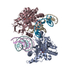 7v0wC  4lezS S: Starting model for refinement C: citing same article ( |
|---|---|
| Similar structure data |
-Links
 PDBj
PDBj


- Assembly
| Deposited unit | 
| ||||||||
|---|---|---|---|---|---|---|---|---|---|
| 1 |
| ||||||||
| Unit cell |
|
-Components
-Protein / DNA chain , 2 types, 6 molecules ACEFIJ
| #1: Protein | Mass: 42638.281 Da / Num. of mol.: 2 / Mutation: E211Q, D213N Source method: isolated from a genetically manipulated source Source: (gene. exp.) Details (production host): His*6-MBP-Tev-AgeI-mcGAS CAT, Kanamycin resistance Production host:  #2: DNA chain | Mass: 5514.603 Da / Num. of mol.: 4 / Source method: isolated from a natural source / Source: (natural) DNA molecule (others) / Plasmid details: ordered from IDT |
|---|
-Non-polymers , 4 types, 55 molecules 


| #3: Chemical | ChemComp-GTP /  Mass: 523.180 Da / Num. of mol.: 4 / Source method: obtained synthetically / Formula: C10H16N5O14P3 / Feature type: SUBJECT OF INVESTIGATION / Comment: GTP, energy-carrying molecule*YM Mass: 523.180 Da / Num. of mol.: 4 / Source method: obtained synthetically / Formula: C10H16N5O14P3 / Feature type: SUBJECT OF INVESTIGATION / Comment: GTP, energy-carrying molecule*YM#4: Chemical |  Mass: 24.305 Da / Num. of mol.: 2 / Source method: obtained synthetically / Formula: Mg / Feature type: SUBJECT OF INVESTIGATION Mass: 24.305 Da / Num. of mol.: 2 / Source method: obtained synthetically / Formula: Mg / Feature type: SUBJECT OF INVESTIGATION#5: Chemical |  Mass: 65.409 Da / Num. of mol.: 2 / Source method: obtained synthetically / Formula: Zn Mass: 65.409 Da / Num. of mol.: 2 / Source method: obtained synthetically / Formula: Zn#6: Water | ChemComp-HOH / | Mass: 18.015 Da / Num. of mol.: 47 / Source method: isolated from a natural source / Formula: H2O |
|---|
-Details
| Has ligand of interest | Y |
|---|
-Experimental details
-Experiment
| Experiment | Method: X-RAY DIFFRACTION / Number of used crystals: 1 |
|---|
- Sample preparation
| Crystal | Density Matthews: 2.56 Å3/Da / Density % sol: 51.96 % |
|---|---|
| Crystal grow | Temperature: 277.15 K / Method: vapor diffusion, hanging drop / pH: 6.5 Details: 0.2 M ammonium acetate, 32% MPD, with 0.1 M Bis-Tris pH 6.5 PH range: 6.0-7.0 / Temp details: 4-degree Celsius in cold room |
-Data collection
| Diffraction | Mean temperature: 100 K / Ambient temp details: nitrogen gas stream / Serial crystal experiment: N | ||||||||||||||||||||||||||||||
|---|---|---|---|---|---|---|---|---|---|---|---|---|---|---|---|---|---|---|---|---|---|---|---|---|---|---|---|---|---|---|---|
| Diffraction source | Source: SYNCHROTRON / Site: NSLS-II / Beamline: 17-ID-2 / Wavelength: 0.9793 Å | ||||||||||||||||||||||||||||||
| Detector | Type: DECTRIS EIGER X 16M / Detector: PIXEL / Date: Oct 19, 2021 | ||||||||||||||||||||||||||||||
| Radiation | Monochromator: horizontal bounce Si(111) double crystal monochromator Protocol: SINGLE WAVELENGTH / Monochromatic (M) / Laue (L): M / Scattering type: x-ray | ||||||||||||||||||||||||||||||
| Radiation wavelength | Wavelength: 0.9793 Å / Relative weight: 1 | ||||||||||||||||||||||||||||||
| Reflection | Resolution: 2.57→28.92 Å / Num. obs: 35696 / % possible obs: 99.4 % / Redundancy: 6.7 % / CC1/2: 0.998 / Rmerge(I) obs: 0.09 / Rpim(I) all: 0.038 / Rrim(I) all: 0.098 / Net I/σ(I): 12.8 | ||||||||||||||||||||||||||||||
| Reflection shell | Diffraction-ID: 1
|
- Processing
| Software |
| |||||||||||||||||||||||||||||||||||||||||||||||||||||||||||||||||||||||||||||||||||||||||||||||||||||||||||||||||||||||||||||||||||||||||||||||||||||||||||||||||||||||||||||||
|---|---|---|---|---|---|---|---|---|---|---|---|---|---|---|---|---|---|---|---|---|---|---|---|---|---|---|---|---|---|---|---|---|---|---|---|---|---|---|---|---|---|---|---|---|---|---|---|---|---|---|---|---|---|---|---|---|---|---|---|---|---|---|---|---|---|---|---|---|---|---|---|---|---|---|---|---|---|---|---|---|---|---|---|---|---|---|---|---|---|---|---|---|---|---|---|---|---|---|---|---|---|---|---|---|---|---|---|---|---|---|---|---|---|---|---|---|---|---|---|---|---|---|---|---|---|---|---|---|---|---|---|---|---|---|---|---|---|---|---|---|---|---|---|---|---|---|---|---|---|---|---|---|---|---|---|---|---|---|---|---|---|---|---|---|---|---|---|---|---|---|---|---|---|---|---|---|
| Refinement | Method to determine structure: MOLECULAR REPLACEMENT Starting model: 4LEZ Resolution: 2.57→28.92 Å / Cor.coef. Fo:Fc: 0.956 / Cor.coef. Fo:Fc free: 0.934 / SU B: 22.048 / SU ML: 0.236 / Cross valid method: THROUGHOUT / ESU R: 0.651 / ESU R Free: 0.294 / Stereochemistry target values: MAXIMUM LIKELIHOOD Details: U VALUES : WITH TLS ADDED HYDROGENS HAVE BEEN ADDED IN THE RIDING POSITIONS U VALUES : RESIDUAL ONLY
| |||||||||||||||||||||||||||||||||||||||||||||||||||||||||||||||||||||||||||||||||||||||||||||||||||||||||||||||||||||||||||||||||||||||||||||||||||||||||||||||||||||||||||||||
| Solvent computation | Ion probe radii: 0.8 Å / Shrinkage radii: 0.8 Å / VDW probe radii: 1.1 Å / Solvent model: MASK | |||||||||||||||||||||||||||||||||||||||||||||||||||||||||||||||||||||||||||||||||||||||||||||||||||||||||||||||||||||||||||||||||||||||||||||||||||||||||||||||||||||||||||||||
| Displacement parameters | Biso max: 191.4 Å2 / Biso mean: 65.737 Å2 / Biso min: 27.66 Å2
| |||||||||||||||||||||||||||||||||||||||||||||||||||||||||||||||||||||||||||||||||||||||||||||||||||||||||||||||||||||||||||||||||||||||||||||||||||||||||||||||||||||||||||||||
| Refinement step | Cycle: final / Resolution: 2.57→28.92 Å
| |||||||||||||||||||||||||||||||||||||||||||||||||||||||||||||||||||||||||||||||||||||||||||||||||||||||||||||||||||||||||||||||||||||||||||||||||||||||||||||||||||||||||||||||
| Refine LS restraints |
| |||||||||||||||||||||||||||||||||||||||||||||||||||||||||||||||||||||||||||||||||||||||||||||||||||||||||||||||||||||||||||||||||||||||||||||||||||||||||||||||||||||||||||||||
| LS refinement shell | Resolution: 2.571→2.638 Å / Rfactor Rfree error: 0 / Total num. of bins used: 20
| |||||||||||||||||||||||||||||||||||||||||||||||||||||||||||||||||||||||||||||||||||||||||||||||||||||||||||||||||||||||||||||||||||||||||||||||||||||||||||||||||||||||||||||||
| Refinement TLS params. | Method: refined / Refine-ID: X-RAY DIFFRACTION
| |||||||||||||||||||||||||||||||||||||||||||||||||||||||||||||||||||||||||||||||||||||||||||||||||||||||||||||||||||||||||||||||||||||||||||||||||||||||||||||||||||||||||||||||
| Refinement TLS group |
|
