 Movie
Movie Controller

[English] 日本語
 Yorodumi
- PDB-7uxd: Crystal structure of APOBEC3G Catalytic domain complex with ssDNA... -
+ Open data

- Basic information
| Entry | Database: PDB / ID: 7uxd | |||||||||
|---|---|---|---|---|---|---|---|---|---|---|
| Title | Crystal structure of APOBEC3G Catalytic domain complex with ssDNA containing 2'-deoxy Zebularine. | |||||||||
 Components Components |
| |||||||||
 Keywords Keywords | HYDROLASE/DNA / DNA CYTIDINE DEAMINASE / APOBEC / APOBEC- inhibitor COMPLEX / Zebularine / HYDROLASE-DNA complex | |||||||||
| Function / homology |  Function and homology information Function and homology informationapolipoprotein B mRNA editing enzyme complex / dCTP deaminase activity / : / single-stranded DNA cytosine deaminase / base conversion or substitution editing / negative regulation of single stranded viral RNA replication via double stranded DNA intermediate / DNA cytosine deamination / cytidine to uridine editing / negative regulation of viral process / cytidine deaminase activity ...apolipoprotein B mRNA editing enzyme complex / dCTP deaminase activity / : / single-stranded DNA cytosine deaminase / base conversion or substitution editing / negative regulation of single stranded viral RNA replication via double stranded DNA intermediate / DNA cytosine deamination / cytidine to uridine editing / negative regulation of viral process / cytidine deaminase activity / transposable element silencing / negative regulation of viral genome replication / APOBEC3G mediated resistance to HIV-1 infection / positive regulation of defense response to virus by host / P-body / Vif-mediated degradation of APOBEC3G / defense response to virus / ribonucleoprotein complex / innate immune response / RNA binding / zinc ion binding / identical protein binding / nucleus / cytoplasm / cytosol Similarity search - Function | |||||||||
| Biological species |  Homo sapiens (human) Homo sapiens (human)synthetic construct (others) | |||||||||
| Method |  X-RAY DIFFRACTION / SYNCHROTRON / MOLECULAR REPLACEMENT / Resolution: 1.5 Å X-RAY DIFFRACTION / SYNCHROTRON / MOLECULAR REPLACEMENT / Resolution: 1.5 Å | |||||||||
 Authors Authors | Maiti, A. / Matsuo, H. | |||||||||
| Funding support |  United States, 2items United States, 2items
| |||||||||
 Citation Citation | Journal: Nat Commun / Year: 2022 Title: Structure of the catalytically active APOBEC3G bound to a DNA oligonucleotide inhibitor reveals tetrahedral geometry of the transition state. Authors: Maiti, A. / Hedger, A.K. / Myint, W. / Balachandran, V. / Watts, J.K. / Schiffer, C.A. / Matsuo, H. | |||||||||
| History |
|
- Structure visualization
| Structure viewer | Molecule: MolmilJmol/JSmol |
|---|
- Downloads & links
-Download
| PDBx/mmCIF format | 7uxd.cif.gz | 117.1 KB | Display | PDBx/mmCIF format |
|---|---|---|---|---|
| PDB format | pdb7uxd.ent.gz | 84.6 KB | Display | PDB format |
| PDBx/mmJSON format | 7uxd.json.gz | Tree view | PDBx/mmJSON format | |
| Others |  Other downloads Other downloads |
-Validation report
| Arichive directory | https://data.pdbj.org/pub/pdb/validation_reports/ux/7uxdftp://data.pdbj.org/pub/pdb/validation_reports/ux/7uxd | HTTPS FTP |
|---|
-Related structure data
| Related structure data | 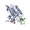 6buxS S: Starting model for refinement |
|---|---|
| Similar structure data |
-Links
 PDBj
PDBj


- Assembly
| Deposited unit | 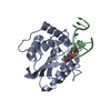
| ||||||||
|---|---|---|---|---|---|---|---|---|---|
| 1 |
| ||||||||
| Unit cell |
|
-Components
| #1: Protein | Mass: 23120.125 Da / Num. of mol.: 1 Mutation: P200A, L234K, N236A, C243A, P247K, F310K, Q318K, C321A, Q322A, C356A Source method: isolated from a genetically manipulated source Source: (gene. exp.) Homo sapiens (human) / Gene: APOBEC3G, MDS019 / Variant: UNP Residues 191-384 / Production host:  References: UniProt: Q9HC16, single-stranded DNA cytosine deaminase |
|---|---|
| #2: DNA chain | Mass: 2695.816 Da / Num. of mol.: 1 Source method: isolated from a genetically manipulated source Source: (gene. exp.) synthetic construct (others) / Production host: synthetic construct (others) |
| #3: Chemical | ChemComp-ZN /   Mass: 65.409 Da / Num. of mol.: 1 / Source method: obtained synthetically / Formula: Zn / Feature type: SUBJECT OF INVESTIGATION Mass: 65.409 Da / Num. of mol.: 1 / Source method: obtained synthetically / Formula: Zn / Feature type: SUBJECT OF INVESTIGATION |
| #4: Water | ChemComp-HOH /  Mass: 18.015 Da / Num. of mol.: 212 / Source method: isolated from a natural source / Formula: H2O Mass: 18.015 Da / Num. of mol.: 212 / Source method: isolated from a natural source / Formula: H2O |
| Has ligand of interest | Y |
-Experimental details
-Experiment
| Experiment | Method: X-RAY DIFFRACTION / Number of used crystals: 1 |
|---|
- Sample preparation
| Crystal | Density Matthews: 2.2 Å3/Da / Density % sol: 43.99 % |
|---|---|
| Crystal grow | Temperature: 277 K / Method: vapor diffusion, sitting drop Details: 20% PEG6000, 50 mM di-Sodium L-Malate; pH5.0, 30 mM CACL2 |
-Data collection
| Diffraction | Mean temperature: 100 K / Serial crystal experiment: N | |||||||||||||||||||||||||||||||||||||||||||||||||||||||||||||||||||||||||||||||||||||||||||||||||||
|---|---|---|---|---|---|---|---|---|---|---|---|---|---|---|---|---|---|---|---|---|---|---|---|---|---|---|---|---|---|---|---|---|---|---|---|---|---|---|---|---|---|---|---|---|---|---|---|---|---|---|---|---|---|---|---|---|---|---|---|---|---|---|---|---|---|---|---|---|---|---|---|---|---|---|---|---|---|---|---|---|---|---|---|---|---|---|---|---|---|---|---|---|---|---|---|---|---|---|---|---|
| Diffraction source | Source: SYNCHROTRON / Site: APS / Beamline: 22-ID / Wavelength: 1 Å | |||||||||||||||||||||||||||||||||||||||||||||||||||||||||||||||||||||||||||||||||||||||||||||||||||
| Detector | Type: DECTRIS EIGER X 16M / Detector: PIXEL / Date: Feb 12, 2022 | |||||||||||||||||||||||||||||||||||||||||||||||||||||||||||||||||||||||||||||||||||||||||||||||||||
| Radiation | Protocol: SINGLE WAVELENGTH / Monochromatic (M) / Laue (L): M / Scattering type: x-ray | |||||||||||||||||||||||||||||||||||||||||||||||||||||||||||||||||||||||||||||||||||||||||||||||||||
| Radiation wavelength | Wavelength: 1 Å / Relative weight: 1 | |||||||||||||||||||||||||||||||||||||||||||||||||||||||||||||||||||||||||||||||||||||||||||||||||||
| Reflection | Resolution: 1.5→50 Å / Num. obs: 33649 / % possible obs: 93.4 % / Redundancy: 3.2 % / Rmerge(I) obs: 0.086 / Rpim(I) all: 0.058 / Rrim(I) all: 0.104 / Χ2: 0.844 / Net I/σ(I): 7.7 / Num. measured all: 106388 | |||||||||||||||||||||||||||||||||||||||||||||||||||||||||||||||||||||||||||||||||||||||||||||||||||
| Reflection shell | Diffraction-ID: 1
|
- Processing
| Software |
| ||||||||||||||||||||||||||||||||||||||||||||||||||||||||||||||||||||||||||||||
|---|---|---|---|---|---|---|---|---|---|---|---|---|---|---|---|---|---|---|---|---|---|---|---|---|---|---|---|---|---|---|---|---|---|---|---|---|---|---|---|---|---|---|---|---|---|---|---|---|---|---|---|---|---|---|---|---|---|---|---|---|---|---|---|---|---|---|---|---|---|---|---|---|---|---|---|---|---|---|---|
| Refinement | Method to determine structure: MOLECULAR REPLACEMENT Starting model: 6BUX (Chain A) Resolution: 1.5→34.506 Å / SU ML: 0.15 / Cross valid method: THROUGHOUT / σ(F): 1.39 / Phase error: 17.56 / Stereochemistry target values: ML
| ||||||||||||||||||||||||||||||||||||||||||||||||||||||||||||||||||||||||||||||
| Solvent computation | Shrinkage radii: 0.9 Å / VDW probe radii: 1.11 Å / Solvent model: FLAT BULK SOLVENT MODEL | ||||||||||||||||||||||||||||||||||||||||||||||||||||||||||||||||||||||||||||||
| Displacement parameters | Biso max: 94.38 Å2 / Biso mean: 36.7548 Å2 / Biso min: 15.57 Å2 | ||||||||||||||||||||||||||||||||||||||||||||||||||||||||||||||||||||||||||||||
| Refinement step | Cycle: final / Resolution: 1.5→34.506 Å
| ||||||||||||||||||||||||||||||||||||||||||||||||||||||||||||||||||||||||||||||
| LS refinement shell | Refine-ID: X-RAY DIFFRACTION / Rfactor Rfree error: 0
| ||||||||||||||||||||||||||||||||||||||||||||||||||||||||||||||||||||||||||||||
| Refinement TLS params. | Method: refined / Origin x: 10.3286 Å / Origin y: -4.9278 Å / Origin z: 13.0935 Å
| ||||||||||||||||||||||||||||||||||||||||||||||||||||||||||||||||||||||||||||||
| Refinement TLS group |
|
