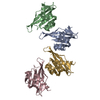
- PDB-7urz: Hexadecameric hub domain of CaMKII beta -
+
Open data
ID or keywords:
Loading...
-
Basic information
Entry
Database: PDB / ID: 7urz
Title
Hexadecameric hub domain of CaMKII beta
Components
Calcium/calmodulin-dependent protein kinase type II subunit beta
Keywords
TRANSFERASE / CaMKII / Kinase / Human / CAMK2B
Function / homology
Function and homology information
regulation of skeletal muscle adaptation / regulation of synapse structural plasticity / calcium- and calmodulin-dependent protein kinase complex / positive regulation of synapse maturation / regulation of dendritic spine development / Ca2+/calmodulin-dependent protein kinase / Trafficking of AMPA receptors / positive regulation of dendritic spine morphogenesis / regulation of neuron migration / calcium/calmodulin-dependent protein kinase activity ...regulation of skeletal muscle adaptation / regulation of synapse structural plasticity / calcium- and calmodulin-dependent protein kinase complex / positive regulation of synapse maturation / regulation of dendritic spine development / Ca2+/calmodulin-dependent protein kinase / Trafficking of AMPA receptors / positive regulation of dendritic spine morphogenesis / regulation of neuron migration / calcium/calmodulin-dependent protein kinase activity / Assembly and cell surface presentation of NMDA receptors / CREB1 phosphorylation through the activation of CaMKII/CaMKK/CaMKIV cascasde / CaMK IV-mediated phosphorylation of CREB / Phase 0 - rapid depolarisation / regulation of calcium ion transport / Negative regulation of NMDA receptor-mediated neuronal transmission / Unblocking of NMDA receptors, glutamate binding and activation / Ion transport by P-type ATPases / Long-term potentiation / HSF1-dependent transactivation / Regulation of MECP2 expression and activity / regulation of neuronal synaptic plasticity / regulation of protein localization to plasma membrane / Ion homeostasis / sarcoplasmic reticulum membrane / Ras activation upon Ca2+ influx through NMDA receptor / RAF activation / positive regulation of neuron projection development / regulation of long-term neuronal synaptic plasticity / Interferon gamma signaling / Signaling by RAF1 mutants / Signaling by moderate kinase activity BRAF mutants / Paradoxical activation of RAF signaling by kinase inactive BRAF / Signaling downstream of RAS mutants / endocytic vesicle membrane / Signaling by BRAF and RAF1 fusions / long-term synaptic potentiation / protein autophosphorylation / nervous system development / RAF/MAP kinase cascade / actin binding / protein phosphorylation / cell differentiation / calmodulin binding / neuron projection / postsynaptic density / protein serine kinase activity / centrosome / signal transduction / protein homodimerization activity / nucleoplasm / ATP binding / identical protein binding / cytoplasm / cytosol Similarity search - Function
Calcium/calmodulin-dependent protein kinase II, association-domain / Calcium/calmodulin dependent protein kinase II association domain / NTF2-like domain superfamily / Serine/threonine-protein kinase, active site / Serine/Threonine protein kinases active-site signature. / Protein kinase domain / Serine/Threonine protein kinases, catalytic domain / Protein kinase, ATP binding site / Protein kinases ATP-binding region signature. / Protein kinase domain profile. ...Calcium/calmodulin-dependent protein kinase II, association-domain / Calcium/calmodulin dependent protein kinase II association domain / NTF2-like domain superfamily / Serine/threonine-protein kinase, active site / Serine/Threonine protein kinases active-site signature. / Protein kinase domain / Serine/Threonine protein kinases, catalytic domain / Protein kinase, ATP binding site / Protein kinases ATP-binding region signature. / Protein kinase domain profile. / Protein kinase domain / Protein kinase-like domain superfamily Similarity search - Domain/homology
G: Calcium/calmodulin-dependent protein kinase type II subunit beta A: Calcium/calmodulin-dependent protein kinase type II subunit beta B: Calcium/calmodulin-dependent protein kinase type II subunit beta C: Calcium/calmodulin-dependent protein kinase type II subunit beta
G: Calcium/calmodulin-dependent protein kinase type II subunit beta A: Calcium/calmodulin-dependent protein kinase type II subunit beta B: Calcium/calmodulin-dependent protein kinase type II subunit beta C: Calcium/calmodulin-dependent protein kinase type II subunit beta
G: Calcium/calmodulin-dependent protein kinase type II subunit beta A: Calcium/calmodulin-dependent protein kinase type II subunit beta B: Calcium/calmodulin-dependent protein kinase type II subunit beta C: Calcium/calmodulin-dependent protein kinase type II subunit beta
G: Calcium/calmodulin-dependent protein kinase type II subunit beta A: Calcium/calmodulin-dependent protein kinase type II subunit beta B: Calcium/calmodulin-dependent protein kinase type II subunit beta C: Calcium/calmodulin-dependent protein kinase type II subunit beta
G: Calcium/calmodulin-dependent protein kinase type II subunit beta A: Calcium/calmodulin-dependent protein kinase type II subunit beta B: Calcium/calmodulin-dependent protein kinase type II subunit beta C: Calcium/calmodulin-dependent protein kinase type II subunit beta
Resolution: 3.45→37.09 Å / Cor.coef. Fo:Fc: 0.812 / Cor.coef. Fo:Fc free: 0.712 / SU B: 61.406 / SU ML: 0.901 / Cross valid method: THROUGHOUT / σ(F): 0 / ESU R Free: 1.012 / Stereochemistry target values: MAXIMUM LIKELIHOOD Details: HYDROGENS HAVE BEEN ADDED IN THE RIDING POSITIONS U VALUES : REFINED INDIVIDUALLY
Rfactor
Num. reflection
% reflection
Selection details
Rfree
0.4074
424
5.1 %
RANDOM
Rwork
0.3516
-
-
-
obs
0.3544
7921
97.75 %
-
Solvent computation
Ion probe radii: 0.8 Å / Shrinkage radii: 0.8 Å / VDW probe radii: 1.2 Å / Solvent model: MASK
In the structure databanks used in Yorodumi, some data are registered as the other names, "COVID-19 virus" and "2019-nCoV". Here are the details of the virus and the list of structure data.
Jan 31, 2019. EMDB accession codes are about to change! (news from PDBe EMDB page)
EMDB accession codes are about to change! (news from PDBe EMDB page)
The allocation of 4 digits for EMDB accession codes will soon come to an end. Whilst these codes will remain in use, new EMDB accession codes will include an additional digit and will expand incrementally as the available range of codes is exhausted. The current 4-digit format prefixed with “EMD-” (i.e. EMD-XXXX) will advance to a 5-digit format (i.e. EMD-XXXXX), and so on. It is currently estimated that the 4-digit codes will be depleted around Spring 2019, at which point the 5-digit format will come into force.
The EM Navigator/Yorodumi systems omit the EMD- prefix.
Related info.:Q: What is EMD? / ID/Accession-code notation in Yorodumi/EM Navigator
Yorodumi is a browser for structure data from EMDB, PDB, SASBDB, etc.
This page is also the successor to EM Navigator detail page, and also detail information page/front-end page for Omokage search.
The word "yorodu" (or yorozu) is an old Japanese word meaning "ten thousand". "mi" (miru) is to see.
Related info.:EMDB / PDB / SASBDB / Comparison of 3 databanks / Yorodumi Search / Aug 31, 2016. New EM Navigator & Yorodumi / Yorodumi Papers / Jmol/JSmol / Function and homology information / Changes in new EM Navigator and Yorodumi
 Movie
Movie Controller


 Open data
 Basic information
 Components
 Keywords
 Function and homology information
 Homo sapiens (human)
 X-RAY DIFFRACTION /
  Authors
 United States, 1items
  Citation
 Structure visualization
 Downloads & links
 Other downloads
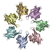
 PDBj
PDBj


 Assembly

 Sample preparation
 Processing