 Movie
Movie Controller

[English] 日本語
 Yorodumi
- PDB-7urs: Crystal Structure of SARS-CoV-2 S delta variant receptor-binding ... -
+ Open data

- Basic information
| Entry | Database: PDB / ID: 7urs | ||||||||||||
|---|---|---|---|---|---|---|---|---|---|---|---|---|---|
| Title | Crystal Structure of SARS-CoV-2 S delta variant receptor-binding domain (RBD) in complex CoV11 Fab crystal form 2 | ||||||||||||
 Components Components |
| ||||||||||||
 Keywords Keywords | VIRAL PROTEIN/Immune System / SARS-CoV-2 S RBD Fab complex / SARS-CoV-2 S receptor-binding domain antibody Fab complex / COVID-19 spike protein antibody Fab complex / delta variant / VIRAL PROTEIN-Immune System complex | ||||||||||||
| Function / homology |  Function and homology information Function and homology informationsymbiont-mediated disruption of host tissue / Maturation of spike protein / Translation of Structural Proteins / Virion Assembly and Release / host cell surface / viral translation / host extracellular space / symbiont-mediated-mediated suppression of host tetherin activity / Induction of Cell-Cell Fusion / structural constituent of virion ...symbiont-mediated disruption of host tissue / Maturation of spike protein / Translation of Structural Proteins / Virion Assembly and Release / host cell surface / viral translation / host extracellular space / symbiont-mediated-mediated suppression of host tetherin activity / Induction of Cell-Cell Fusion / structural constituent of virion / entry receptor-mediated virion attachment to host cell / membrane fusion / Attachment and Entry / host cell endoplasmic reticulum-Golgi intermediate compartment membrane / positive regulation of viral entry into host cell / receptor-mediated virion attachment to host cell / host cell surface receptor binding / symbiont-mediated suppression of host innate immune response / receptor ligand activity / endocytosis involved in viral entry into host cell / fusion of virus membrane with host plasma membrane / fusion of virus membrane with host endosome membrane / viral envelope / symbiont entry into host cell / virion attachment to host cell / SARS-CoV-2 activates/modulates innate and adaptive immune responses / host cell plasma membrane / virion membrane / identical protein binding / membrane / plasma membrane Similarity search - Function | ||||||||||||
| Biological species |   Severe acute respiratory syndrome coronavirus 2 Severe acute respiratory syndrome coronavirus 2 Homo sapiens (human) Homo sapiens (human) | ||||||||||||
| Method |  X-RAY DIFFRACTION / SYNCHROTRON / MOLECULAR REPLACEMENT / Resolution: 2.4 Å X-RAY DIFFRACTION / SYNCHROTRON / MOLECULAR REPLACEMENT / Resolution: 2.4 Å | ||||||||||||
 Authors Authors | Tolbert, W.D. / Pazgier, M. | ||||||||||||
| Funding support |  United States, 3items United States, 3items
| ||||||||||||
 Citation Citation | Journal: Front Immunol / Year: 2023 Title: The molecular basis of the neutralization breadth of the RBD-specific antibody CoV11. Authors: Tolbert, W.D. / Chen, Y. / Sun, L. / Benlarbi, M. / Ding, S. / Manickam, R. / Pangaro, E. / Nguyen, D.N. / Gottumukkala, S. / Cote, M. / Gonzalez, F.J. / Finzi, A. / Tehrani, Z.R. / Sajadi, M.M. / Pazgier, M. | ||||||||||||
| History |
|
- Structure visualization
| Structure viewer | Molecule: MolmilJmol/JSmol |
|---|
- Downloads & links
-Download
| PDBx/mmCIF format | 7urs.cif.gz | 259 KB | Display | PDBx/mmCIF format |
|---|---|---|---|---|
| PDB format | pdb7urs.ent.gz | 205.9 KB | Display | PDB format |
| PDBx/mmJSON format | 7urs.json.gz | Tree view | PDBx/mmJSON format | |
| Others |  Other downloads Other downloads |
-Validation report
| Summary document | 7urs_validation.pdf.gz | 482.9 KB | Display | wwPDB validaton report |
|---|---|---|---|---|
| Full document | 7urs_full_validation.pdf.gz | 491.5 KB | Display | |
| Data in XML | 7urs_validation.xml.gz | 24 KB | Display | |
| Data in CIF | 7urs_validation.cif.gz | 33.6 KB | Display | |
| Arichive directory | https://data.pdbj.org/pub/pdb/validation_reports/ur/7ursftp://data.pdbj.org/pub/pdb/validation_reports/ur/7urs | HTTPS FTP |
-Related structure data
| Related structure data | 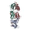 7s4sSC 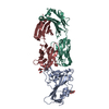 7urqC S: Starting model for refinement C: citing same article ( |
|---|---|
| Similar structure data |
-Links
 PDBj
PDBj


- Assembly
| Deposited unit | 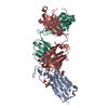
| ||||||||
|---|---|---|---|---|---|---|---|---|---|
| 1 |
| ||||||||
| Unit cell |
|
-Components
-Antibody , 2 types, 2 molecules HL
| #2: Antibody | Mass: 23362.287 Da / Num. of mol.: 1 Source method: isolated from a genetically manipulated source Source: (gene. exp.) Homo sapiens (human) / Cell (production host): HEK 293 / Production host: Homo sapiens (human) |
|---|---|
| #3: Antibody | Mass: 23536.008 Da / Num. of mol.: 1 Source method: isolated from a genetically manipulated source Source: (gene. exp.) Homo sapiens (human) / Cell (production host): HEK 293 / Production host: Homo sapiens (human) |
-Protein / Sugars , 2 types, 2 molecules A
| #1: Protein | Mass: 26167.459 Da / Num. of mol.: 1 / Mutation: L452R, T478K Source method: isolated from a genetically manipulated source Source: (gene. exp.) Severe acute respiratory syndrome coronavirus 2Gene: S, 2 / Cell (production host): HEK 293 GnT1- / Production host: Homo sapiens (human) / References: UniProt: P0DTC2 |
|---|---|
| #4: Sugar | ChemComp-NAG /  Type: D-saccharide, beta linking / Mass: 221.208 Da / Num. of mol.: 1 Type: D-saccharide, beta linking / Mass: 221.208 Da / Num. of mol.: 1Source method: isolated from a genetically manipulated source Formula: C8H15NO6 |
-Non-polymers , 3 types, 89 molecules 


| #5: Chemical |  Mass: 94.971 Da / Num. of mol.: 2 / Source method: obtained synthetically / Formula: PO4 Mass: 94.971 Da / Num. of mol.: 2 / Source method: obtained synthetically / Formula: PO4#6: Chemical |  Mass: 118.174 Da / Num. of mol.: 2 / Source method: obtained synthetically / Formula: C6H14O2 / Comment: precipitant*YM Mass: 118.174 Da / Num. of mol.: 2 / Source method: obtained synthetically / Formula: C6H14O2 / Comment: precipitant*YM#7: Water | ChemComp-HOH / | Mass: 18.015 Da / Num. of mol.: 85 / Source method: isolated from a natural source / Formula: H2O |
|---|
-Details
| Has ligand of interest | N |
|---|---|
| Has protein modification | Y |
-Experimental details
-Experiment
| Experiment | Method: X-RAY DIFFRACTION / Number of used crystals: 1 |
|---|
- Sample preparation
| Crystal | Density Matthews: 3.28 Å3/Da / Density % sol: 62.47 % |
|---|---|
| Crystal grow | Temperature: 294 K / Method: vapor diffusion, hanging drop / pH: 6.5 / Details: 12% PEG 8000 0.1 M sodium phosphate pH 6.5 |
-Data collection
| Diffraction | Mean temperature: 100 K / Serial crystal experiment: N |
|---|---|
| Diffraction source | Source: SYNCHROTRON / Site: SSRL / Beamline: BL12-2 / Wavelength: 0.9795 Å |
| Detector | Type: DECTRIS PILATUS 6M / Detector: PIXEL / Date: Mar 18, 2022 |
| Radiation | Monochromator: Si (1 1 1) / Protocol: SINGLE WAVELENGTH / Monochromatic (M) / Laue (L): M / Scattering type: x-ray |
| Radiation wavelength | Wavelength: 0.9795 Å / Relative weight: 1 |
| Reflection | Resolution: 2.4→50 Å / Num. obs: 34224 / % possible obs: 92.3 % / Redundancy: 3 % / CC1/2: 0.97 / Rmerge(I) obs: 0.126 / Rpim(I) all: 0.081 / Net I/σ(I): 17.5 |
| Reflection shell | Resolution: 2.4→2.44 Å / Redundancy: 1.9 % / Rmerge(I) obs: 0.38 / Mean I/σ(I) obs: 2.05 / Num. unique obs: 1302 / CC1/2: 0.803 / Rpim(I) all: 0.294 / % possible all: 72 |
- Processing
| Software |
| ||||||||||||||||||||||||||||||||||||||||||||||||||||||||||||||||||||||||||||||||||||||||||||||||||||
|---|---|---|---|---|---|---|---|---|---|---|---|---|---|---|---|---|---|---|---|---|---|---|---|---|---|---|---|---|---|---|---|---|---|---|---|---|---|---|---|---|---|---|---|---|---|---|---|---|---|---|---|---|---|---|---|---|---|---|---|---|---|---|---|---|---|---|---|---|---|---|---|---|---|---|---|---|---|---|---|---|---|---|---|---|---|---|---|---|---|---|---|---|---|---|---|---|---|---|---|---|---|
| Refinement | Method to determine structure: MOLECULAR REPLACEMENT Starting model: 7S4S Resolution: 2.4→41.05 Å / SU ML: 0.32 / Cross valid method: FREE R-VALUE / σ(F): 1.36 / Phase error: 25.86 / Stereochemistry target values: ML
| ||||||||||||||||||||||||||||||||||||||||||||||||||||||||||||||||||||||||||||||||||||||||||||||||||||
| Solvent computation | Shrinkage radii: 0.9 Å / VDW probe radii: 1.11 Å / Solvent model: FLAT BULK SOLVENT MODEL | ||||||||||||||||||||||||||||||||||||||||||||||||||||||||||||||||||||||||||||||||||||||||||||||||||||
| Refinement step | Cycle: LAST / Resolution: 2.4→41.05 Å
| ||||||||||||||||||||||||||||||||||||||||||||||||||||||||||||||||||||||||||||||||||||||||||||||||||||
| Refine LS restraints |
| ||||||||||||||||||||||||||||||||||||||||||||||||||||||||||||||||||||||||||||||||||||||||||||||||||||
| LS refinement shell |
| ||||||||||||||||||||||||||||||||||||||||||||||||||||||||||||||||||||||||||||||||||||||||||||||||||||
| Refinement TLS params. | Method: refined / Refine-ID: X-RAY DIFFRACTION
| ||||||||||||||||||||||||||||||||||||||||||||||||||||||||||||||||||||||||||||||||||||||||||||||||||||
| Refinement TLS group |
|
