 Movie
Movie Controller


+ Open data

- Basic information
| Entry | Database: PDB / ID: 7ur2 | ||||||||||||
|---|---|---|---|---|---|---|---|---|---|---|---|---|---|
| Title | Crystal structure of the Sec14 domain of the RhoGEF Kalirin | ||||||||||||
 Components Components | Isoform 7 of Kalirin | ||||||||||||
 Keywords Keywords | LIPID BINDING PROTEIN / Ras homologous guanine nucleotide exchange factor / CRAL-TRIO domain / spectrin repeat / lysophospholipids | ||||||||||||
| Function / homology |  Function and homology information Function and homology informationEPHB-mediated forward signaling / NRAGE signals death through JNK / maternal process involved in parturition / RAC1 GTPase cycle / G alpha (12/13) signalling events / G alpha (q) signalling events / negative regulation of growth hormone secretion / RHOA GTPase cycle / RHOG GTPase cycle / MAPK6/MAPK4 signaling ...EPHB-mediated forward signaling / NRAGE signals death through JNK / maternal process involved in parturition / RAC1 GTPase cycle / G alpha (12/13) signalling events / G alpha (q) signalling events / negative regulation of growth hormone secretion / RHOA GTPase cycle / RHOG GTPase cycle / MAPK6/MAPK4 signaling / habituation / positive regulation of dendritic spine morphogenesis / maternal behavior / neuromuscular junction development / social behavior / lactation / guanyl-nucleotide exchange factor activity / adult locomotory behavior / memory / cytoskeleton / non-specific serine/threonine protein kinase / protein serine kinase activity / protein serine/threonine kinase activity / ATP binding / metal ion binding / cytoplasm Similarity search - Function | ||||||||||||
| Biological species |  | ||||||||||||
| Method |  X-RAY DIFFRACTION / SYNCHROTRON / SAD / Resolution: 1.89 Å X-RAY DIFFRACTION / SYNCHROTRON / SAD / Resolution: 1.89 Å | ||||||||||||
 Authors Authors | Li, Y. / Doukov, T.I. / Hao, B. | ||||||||||||
| Funding support |  United States, 3items United States, 3items
| ||||||||||||
 Citation Citation | Journal: Nat Commun / Year: 2023 Title: Structure of the Sec14 domain of Kalirin reveals a distinct class of lipid-binding module in RhoGEFs. Authors: Li, Y. / Pustovalova, Y. / Doukov, T.I. / Hoch, J.C. / Mains, R.E. / Eipper, B.A. / Hao, B. | ||||||||||||
| History |
|
- Structure visualization
| Structure viewer | Molecule: MolmilJmol/JSmol |
|---|
- Downloads & links
-Download
| PDBx/mmCIF format | 7ur2.cif.gz | 569.3 KB | Display | PDBx/mmCIF format |
|---|---|---|---|---|
| PDB format | pdb7ur2.ent.gz | 473 KB | Display | PDB format |
| PDBx/mmJSON format | 7ur2.json.gz | Tree view | PDBx/mmJSON format | |
| Others |  Other downloads Other downloads |
-Validation report
| Summary document | 7ur2_validation.pdf.gz | 497.9 KB | Display | wwPDB validaton report |
|---|---|---|---|---|
| Full document | 7ur2_full_validation.pdf.gz | 507.2 KB | Display | |
| Data in XML | 7ur2_validation.xml.gz | 63.1 KB | Display | |
| Data in CIF | 7ur2_validation.cif.gz | 92.2 KB | Display | |
| Arichive directory | https://data.pdbj.org/pub/pdb/validation_reports/ur/7ur2ftp://data.pdbj.org/pub/pdb/validation_reports/ur/7ur2 | HTTPS FTP |
-Related structure data
| Similar structure data |
|---|
-Links
 PDBj
PDBj


- Assembly
| Deposited unit | 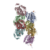
| ||||||||
|---|---|---|---|---|---|---|---|---|---|
| 1 | 
| ||||||||
| 2 | 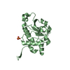
| ||||||||
| 3 | 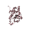
| ||||||||
| 4 | 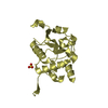
| ||||||||
| 5 | 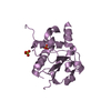
| ||||||||
| 6 | 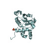
| ||||||||
| 7 | 
| ||||||||
| 8 | 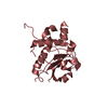
| ||||||||
| Unit cell |
|
-Components
| #1: Protein | Mass: 21742.564 Da / Num. of mol.: 8 / Fragment: Sec14 domain (UNP residues 2-192) Source method: isolated from a genetically manipulated source Source: (gene. exp.)  References: UniProt: A2CG49, non-specific serine/threonine protein kinase #2: Chemical | ChemComp-SO4 /   Mass: 96.063 Da / Num. of mol.: 7 / Source method: obtained synthetically / Formula: SO4 Mass: 96.063 Da / Num. of mol.: 7 / Source method: obtained synthetically / Formula: SO4#3: Water | ChemComp-HOH / |  Mass: 18.015 Da / Num. of mol.: 1412 / Source method: isolated from a natural source / Formula: H2O Mass: 18.015 Da / Num. of mol.: 1412 / Source method: isolated from a natural source / Formula: H2OHas ligand of interest | N | |
|---|
-Experimental details
-Experiment
| Experiment | Method: X-RAY DIFFRACTION / Number of used crystals: 1 |
|---|
- Sample preparation
| Crystal | Density Matthews: 2.65 Å3/Da / Density % sol: 53.54 % |
|---|---|
| Crystal grow | Temperature: 277 K / Method: vapor diffusion, hanging drop / pH: 6.5 Details: 100 mM sodium cacodylate, pH 6.5, 0.2 M ammonium sulfate, 20-25% PEG3350, 0.2 M sodium chloride |
-Data collection
| Diffraction |
| |||||||||||||||||||||||||
|---|---|---|---|---|---|---|---|---|---|---|---|---|---|---|---|---|---|---|---|---|---|---|---|---|---|---|
| Diffraction source |
| |||||||||||||||||||||||||
| Detector |
| |||||||||||||||||||||||||
| Radiation |
| |||||||||||||||||||||||||
| Radiation wavelength |
| |||||||||||||||||||||||||
| Reflection | Resolution: 1.89→78.64 Å / Num. obs: 137673 / % possible obs: 95.9 % / Redundancy: 18.3 % / Biso Wilson estimate: 35.12 Å2 / CC1/2: 1 / Rmerge(I) obs: 0.061 / Rpim(I) all: 0.015 / Rrim(I) all: 0.063 / Net I/σ(I): 31.6 | |||||||||||||||||||||||||
| Reflection shell | Resolution: 1.89→1.92 Å / Redundancy: 19 % / Rmerge(I) obs: 1.294 / Mean I/σ(I) obs: 2.2 / Num. unique obs: 6729 / CC1/2: 0.907 / Rpim(I) all: 0.303 / Rrim(I) all: 1.329 / % possible all: 94 |
- Processing
| Software |
| |||||||||||||||||||||||||||||||||||||||||||||||||||||||||||||||||||||||||||||||||||||||||||||||||||||||||||||||||||||||||||||||||||||||||||||||||||||||||||||||||||||||||||||||||||||||||||||||||||||||||||||||||||||||||||||||||
|---|---|---|---|---|---|---|---|---|---|---|---|---|---|---|---|---|---|---|---|---|---|---|---|---|---|---|---|---|---|---|---|---|---|---|---|---|---|---|---|---|---|---|---|---|---|---|---|---|---|---|---|---|---|---|---|---|---|---|---|---|---|---|---|---|---|---|---|---|---|---|---|---|---|---|---|---|---|---|---|---|---|---|---|---|---|---|---|---|---|---|---|---|---|---|---|---|---|---|---|---|---|---|---|---|---|---|---|---|---|---|---|---|---|---|---|---|---|---|---|---|---|---|---|---|---|---|---|---|---|---|---|---|---|---|---|---|---|---|---|---|---|---|---|---|---|---|---|---|---|---|---|---|---|---|---|---|---|---|---|---|---|---|---|---|---|---|---|---|---|---|---|---|---|---|---|---|---|---|---|---|---|---|---|---|---|---|---|---|---|---|---|---|---|---|---|---|---|---|---|---|---|---|---|---|---|---|---|---|---|---|---|---|---|---|---|---|---|---|---|---|---|---|---|---|---|---|
| Refinement | Method to determine structure: SAD / Resolution: 1.89→61.01 Å / Cor.coef. Fo:Fc: 0.946 / Cor.coef. Fo:Fc free: 0.935 / SU R Cruickshank DPI: 0.138 / Cross valid method: THROUGHOUT / σ(F): 0 / SU R Blow DPI: 0.145 / SU Rfree Blow DPI: 0.127 / SU Rfree Cruickshank DPI: 0.124
| |||||||||||||||||||||||||||||||||||||||||||||||||||||||||||||||||||||||||||||||||||||||||||||||||||||||||||||||||||||||||||||||||||||||||||||||||||||||||||||||||||||||||||||||||||||||||||||||||||||||||||||||||||||||||||||||||
| Displacement parameters | Biso max: 261.83 Å2 / Biso mean: 48.49 Å2 / Biso min: 17.05 Å2
| |||||||||||||||||||||||||||||||||||||||||||||||||||||||||||||||||||||||||||||||||||||||||||||||||||||||||||||||||||||||||||||||||||||||||||||||||||||||||||||||||||||||||||||||||||||||||||||||||||||||||||||||||||||||||||||||||
| Refine analyze | Luzzati coordinate error obs: 0.24 Å | |||||||||||||||||||||||||||||||||||||||||||||||||||||||||||||||||||||||||||||||||||||||||||||||||||||||||||||||||||||||||||||||||||||||||||||||||||||||||||||||||||||||||||||||||||||||||||||||||||||||||||||||||||||||||||||||||
| Refinement step | Cycle: final / Resolution: 1.89→61.01 Å
| |||||||||||||||||||||||||||||||||||||||||||||||||||||||||||||||||||||||||||||||||||||||||||||||||||||||||||||||||||||||||||||||||||||||||||||||||||||||||||||||||||||||||||||||||||||||||||||||||||||||||||||||||||||||||||||||||
| Refine LS restraints |
| |||||||||||||||||||||||||||||||||||||||||||||||||||||||||||||||||||||||||||||||||||||||||||||||||||||||||||||||||||||||||||||||||||||||||||||||||||||||||||||||||||||||||||||||||||||||||||||||||||||||||||||||||||||||||||||||||
| LS refinement shell | Resolution: 1.89→1.9 Å / Rfactor Rfree error: 0 / Total num. of bins used: 50
| |||||||||||||||||||||||||||||||||||||||||||||||||||||||||||||||||||||||||||||||||||||||||||||||||||||||||||||||||||||||||||||||||||||||||||||||||||||||||||||||||||||||||||||||||||||||||||||||||||||||||||||||||||||||||||||||||
| Refinement TLS params. | Method: refined / Refine-ID: X-RAY DIFFRACTION
| |||||||||||||||||||||||||||||||||||||||||||||||||||||||||||||||||||||||||||||||||||||||||||||||||||||||||||||||||||||||||||||||||||||||||||||||||||||||||||||||||||||||||||||||||||||||||||||||||||||||||||||||||||||||||||||||||
| Refinement TLS group |
|
