 Movie
Movie Controller

[English] 日本語
 Yorodumi
- PDB-7uod: Crystal structure of a lectin from Canavalia maritima seed (ConM)... -
+ Open data

- Basic information
| Entry | Database: PDB / ID: 7uod | ||||||
|---|---|---|---|---|---|---|---|
| Title | Crystal structure of a lectin from Canavalia maritima seed (ConM) complexed with 2,4-dichloro-phenoxyacetic acid | ||||||
 Components Components | ConM | ||||||
 Keywords Keywords | SUGAR BINDING PROTEIN / Lectin / Auxin / 2 / 4-Dichlorophenoxyacetic acid | ||||||
| Function / homology | (2,4-DICHLOROPHENOXY)ACETIC ACID / D-ALPHA-AMINOBUTYRIC ACID / : / DI(HYDROXYETHYL)ETHER Function and homology information Function and homology information | ||||||
| Biological species |  Canavalia rosea (plant) Canavalia rosea (plant) | ||||||
| Method |  X-RAY DIFFRACTION / SYNCHROTRON / MOLECULAR REPLACEMENT / Resolution: 1.6 Å X-RAY DIFFRACTION / SYNCHROTRON / MOLECULAR REPLACEMENT / Resolution: 1.6 Å | ||||||
 Authors Authors | Sousa, J.P. / Bezerra, E.H.S. / Sales, M.V. / Queiroz, P.P. / da Silva, F.M.S. / Carvalho, C.P.S. / Freire, V.N. / Rocha, B.A.M. | ||||||
| Funding support |  Brazil, 1items Brazil, 1items
| ||||||
 Citation Citation | Journal: To Be Published Title: Structural analysis of Canavalia maritima lectin complexed with auxins Authors: Sousa, J.P. / Bezerra, E.H.S. / Sales, M.V. / Queiroz, P.P. / da Silva, F.M.S. / Carvalho, C.P.S. / Freire, V.N. / Rocha, B.A.M. | ||||||
| History |
|
- Structure visualization
| Structure viewer | Molecule: MolmilJmol/JSmol |
|---|
- Downloads & links
-Download
| PDBx/mmCIF format | 7uod.cif.gz | 136.8 KB | Display | PDBx/mmCIF format |
|---|---|---|---|---|
| PDB format | pdb7uod.ent.gz | 83.9 KB | Display | PDB format |
| PDBx/mmJSON format | 7uod.json.gz | Tree view | PDBx/mmJSON format | |
| Others |  Other downloads Other downloads |
-Validation report
| Arichive directory | https://data.pdbj.org/pub/pdb/validation_reports/uo/7uodftp://data.pdbj.org/pub/pdb/validation_reports/uo/7uod | HTTPS FTP |
|---|
-Related structure data
| Related structure data | 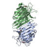 7un2C 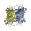 2cy6S S: Starting model for refinement C: citing same article ( |
|---|---|
| Similar structure data |
-Links
 PDBj
PDBj

- Assembly
| Deposited unit | 
| ||||||||||||
|---|---|---|---|---|---|---|---|---|---|---|---|---|---|
| 1 | 
| ||||||||||||
| Unit cell |
|
-Components
-Protein , 1 types, 2 molecules AB
| #1: Protein | Mass: 25494.195 Da / Num. of mol.: 2 / Source method: isolated from a natural source / Source: (natural) Canavalia rosea (plant) |
|---|
-Non-polymers , 8 types, 331 molecules 


| #2: Chemical |  Type: D-peptide linking / Mass: 103.120 Da / Num. of mol.: 2 / Source method: obtained synthetically / Formula: C4H9NO2 / Feature type: SUBJECT OF INVESTIGATION Type: D-peptide linking / Mass: 103.120 Da / Num. of mol.: 2 / Source method: obtained synthetically / Formula: C4H9NO2 / Feature type: SUBJECT OF INVESTIGATION#3: Chemical |  Mass: 40.078 Da / Num. of mol.: 2 / Source method: obtained synthetically / Formula: Ca Mass: 40.078 Da / Num. of mol.: 2 / Source method: obtained synthetically / Formula: Ca#4: Chemical |  Mass: 54.938 Da / Num. of mol.: 2 / Source method: obtained synthetically / Formula: Mn Mass: 54.938 Da / Num. of mol.: 2 / Source method: obtained synthetically / Formula: Mn#5: Chemical |  Mass: 92.094 Da / Num. of mol.: 2 / Source method: obtained synthetically / Formula: C3H8O3 Mass: 92.094 Da / Num. of mol.: 2 / Source method: obtained synthetically / Formula: C3H8O3#6: Chemical |  Mass: 106.120 Da / Num. of mol.: 2 / Source method: obtained synthetically / Formula: C4H10O3 Mass: 106.120 Da / Num. of mol.: 2 / Source method: obtained synthetically / Formula: C4H10O3#7: Chemical | ChemComp-CL /  Mass: 35.453 Da / Num. of mol.: 4 / Source method: obtained synthetically / Formula: Cl Mass: 35.453 Da / Num. of mol.: 4 / Source method: obtained synthetically / Formula: Cl#8: Chemical | ChemComp-CFA / ( |  Mass: 221.037 Da / Num. of mol.: 1 / Source method: obtained synthetically / Formula: C8H6Cl2O3 / Feature type: SUBJECT OF INVESTIGATION Mass: 221.037 Da / Num. of mol.: 1 / Source method: obtained synthetically / Formula: C8H6Cl2O3 / Feature type: SUBJECT OF INVESTIGATION#9: Water | ChemComp-HOH / | Mass: 18.015 Da / Num. of mol.: 316 / Source method: isolated from a natural source / Formula: H2O |
|---|
-Details
| Has ligand of interest | Y |
|---|
-Experimental details
-Experiment
| Experiment | Method: X-RAY DIFFRACTION / Number of used crystals: 1 |
|---|
- Sample preparation
| Crystal | Density Matthews: 2.37 Å3/Da / Density % sol: 48.07 % |
|---|---|
| Crystal grow | Temperature: 298 K / Method: vapor diffusion, hanging drop / pH: 8.5 / Details: 100 mM HEPES, 2.0 M Ammonium sulfate, 4% PEG 400 |
-Data collection
| Diffraction | Mean temperature: 100 K / Serial crystal experiment: N |
|---|---|
| Diffraction source | Source: SYNCHROTRON / Site: LNLS / Beamline: W01B-MX2 / Wavelength: 1.458 Å |
| Detector | Type: DECTRIS PILATUS 2M / Detector: PIXEL / Date: Mar 1, 2018 |
| Radiation | Protocol: SINGLE WAVELENGTH / Monochromatic (M) / Laue (L): M / Scattering type: x-ray |
| Radiation wavelength | Wavelength: 1.458 Å / Relative weight: 1 |
| Reflection | Resolution: 1.6→33.75 Å / Num. obs: 62417 / % possible obs: 93.31 % / Redundancy: 2 % / Biso Wilson estimate: 20.06 Å2 / CC1/2: 1 / Net I/σ(I): 26.14 |
| Reflection shell | Resolution: 1.6→1.657 Å / Mean I/σ(I) obs: 3.44 / Num. unique obs: 5886 / CC1/2: 0.908 / % possible all: 92.26 |
- Processing
| Software |
| |||||||||||||||||||||||||||||||||||||||||||||||||||||||||||||||||||||||||||||||||||||||||||||||||||||||||
|---|---|---|---|---|---|---|---|---|---|---|---|---|---|---|---|---|---|---|---|---|---|---|---|---|---|---|---|---|---|---|---|---|---|---|---|---|---|---|---|---|---|---|---|---|---|---|---|---|---|---|---|---|---|---|---|---|---|---|---|---|---|---|---|---|---|---|---|---|---|---|---|---|---|---|---|---|---|---|---|---|---|---|---|---|---|---|---|---|---|---|---|---|---|---|---|---|---|---|---|---|---|---|---|---|---|---|
| Refinement | Method to determine structure: MOLECULAR REPLACEMENT Starting model: 2CY6 Resolution: 1.6→33.75 Å / SU ML: 0.1818 / Cross valid method: FREE R-VALUE / σ(F): 1.34 / Phase error: 25.1636 Stereochemistry target values: GeoStd + Monomer Library + CDL v1.2
| |||||||||||||||||||||||||||||||||||||||||||||||||||||||||||||||||||||||||||||||||||||||||||||||||||||||||
| Solvent computation | Shrinkage radii: 0.9 Å / VDW probe radii: 1.11 Å / Solvent model: FLAT BULK SOLVENT MODEL | |||||||||||||||||||||||||||||||||||||||||||||||||||||||||||||||||||||||||||||||||||||||||||||||||||||||||
| Displacement parameters | Biso mean: 24.91 Å2 | |||||||||||||||||||||||||||||||||||||||||||||||||||||||||||||||||||||||||||||||||||||||||||||||||||||||||
| Refinement step | Cycle: LAST / Resolution: 1.6→33.75 Å
| |||||||||||||||||||||||||||||||||||||||||||||||||||||||||||||||||||||||||||||||||||||||||||||||||||||||||
| Refine LS restraints |
| |||||||||||||||||||||||||||||||||||||||||||||||||||||||||||||||||||||||||||||||||||||||||||||||||||||||||
| LS refinement shell |
|
