 Movie
Movie Controller


+ Open data

- Basic information
| Entry | Database: PDB / ID: 7uls | ||||||
|---|---|---|---|---|---|---|---|
| Title | Recombinant muscarinic toxin alpha | ||||||
 Components Components | Muscarinic toxin alpha | ||||||
 Keywords Keywords | BIOSYNTHETIC PROTEIN / recombinant / muscarinic toxin / refolded | ||||||
| Function / homology | Snake toxin, conserved site / Snake toxins signature. / : / Snake toxin cobra-type / Snake three-finger toxin / Snake toxin-like superfamily / toxin activity / extracellular region / Muscarinic toxin alpha Function and homology information Function and homology information | ||||||
| Biological species |  Dendroaspis polylepis polylepis (black mamba) Dendroaspis polylepis polylepis (black mamba) | ||||||
| Method |  X-RAY DIFFRACTION / SYNCHROTRON / MOLECULAR REPLACEMENT / Resolution: 1.8 Å X-RAY DIFFRACTION / SYNCHROTRON / MOLECULAR REPLACEMENT / Resolution: 1.8 Å | ||||||
 Authors Authors | Xu, J. / Lei, X. / Chen, L. | ||||||
| Funding support |  United States, 1items United States, 1items
| ||||||
 Citation Citation | Journal: To Be Published Title: recombinant MTalpha at 1.8 Angstroms resolution Authors: Xu, J. / Lei, X. / Chen, L. | ||||||
| History |
|
- Structure visualization
| Structure viewer | Molecule: MolmilJmol/JSmol |
|---|
- Downloads & links
-Download
| PDBx/mmCIF format | 7uls.cif.gz | 34 KB | Display | PDBx/mmCIF format |
|---|---|---|---|---|
| PDB format | pdb7uls.ent.gz | 17.1 KB | Display | PDB format |
| PDBx/mmJSON format | 7uls.json.gz | Tree view | PDBx/mmJSON format | |
| Others |  Other downloads Other downloads |
-Validation report
| Arichive directory | https://data.pdbj.org/pub/pdb/validation_reports/ul/7ulsftp://data.pdbj.org/pub/pdb/validation_reports/ul/7uls | HTTPS FTP |
|---|
-Related structure data
| Related structure data | 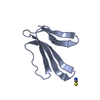 4do8S S: Starting model for refinement |
|---|---|
| Similar structure data |
-Links
 PDBj
PDBj

- Assembly
| Deposited unit | 
| ||||||||||||
|---|---|---|---|---|---|---|---|---|---|---|---|---|---|
| 1 |
| ||||||||||||
| Unit cell |
|
-Components
| #1: Protein | Mass: 7693.773 Da / Num. of mol.: 1 Source method: isolated from a genetically manipulated source Source: (gene. exp.) Dendroaspis polylepis polylepis (black mamba)Production host:  | ||||||
|---|---|---|---|---|---|---|---|
| #2: Chemical |   Mass: 92.094 Da / Num. of mol.: 2 / Source method: obtained synthetically / Formula: C3H8O3 Mass: 92.094 Da / Num. of mol.: 2 / Source method: obtained synthetically / Formula: C3H8O3#3: Water | ChemComp-HOH / |  Mass: 18.015 Da / Num. of mol.: 69 / Source method: isolated from a natural source / Formula: H2O Mass: 18.015 Da / Num. of mol.: 69 / Source method: isolated from a natural source / Formula: H2OHas ligand of interest | N | Has protein modification | Y | |
-Experimental details
-Experiment
| Experiment | Method: X-RAY DIFFRACTION / Number of used crystals: 1 |
|---|
- Sample preparation
| Crystal | Density Matthews: 2.28 Å3/Da / Density % sol: 45.97 % |
|---|---|
| Crystal grow | Temperature: 291.15 K / Method: vapor diffusion, hanging drop Details: 128 mg/ml in 200 mM NH4Ac (pH 7.0), 1:1(v/v) with 1.26 M sodium phosphate monobasic monohydrate, 0.14 M potassium phosphate, pH 5.6 at 18 degree centigrade |
-Data collection
| Diffraction | Mean temperature: 100 K / Serial crystal experiment: N |
|---|---|
| Diffraction source | Source: SYNCHROTRON / Site: APS / Beamline: 19-ID / Wavelength: 1.00007 Å |
| Detector | Type: DECTRIS PILATUS3 X 6M / Detector: PIXEL / Date: Jul 7, 2016 |
| Radiation | Protocol: SINGLE WAVELENGTH / Monochromatic (M) / Laue (L): M / Scattering type: x-ray |
| Radiation wavelength | Wavelength: 1.00007 Å / Relative weight: 1 |
| Reflection | Resolution: 1.5→29.28 Å / Num. obs: 11189 / % possible obs: 99.4 % / Redundancy: 9.3 % / Biso Wilson estimate: 21.26 Å2 / Rpim(I) all: 0.017 / Rrim(I) all: 0.052 / Net I/σ(I): 53.4 |
| Reflection shell | Resolution: 1.5→1.53 Å / Rmerge(I) obs: 0.494 / Num. unique obs: 523 |
- Processing
| Software |
| ||||||||||||||||||||||||
|---|---|---|---|---|---|---|---|---|---|---|---|---|---|---|---|---|---|---|---|---|---|---|---|---|---|
| Refinement | Method to determine structure: MOLECULAR REPLACEMENT Starting model: 4DO8 Resolution: 1.8→29.28 Å / SU ML: 0.0609 / Cross valid method: FREE R-VALUE / σ(F): 1.45 / Phase error: 30.8048 Stereochemistry target values: GeoStd + Monomer Library + CDL v1.2
| ||||||||||||||||||||||||
| Solvent computation | Shrinkage radii: 0.9 Å / VDW probe radii: 1.11 Å / Solvent model: FLAT BULK SOLVENT MODEL | ||||||||||||||||||||||||
| Displacement parameters | Biso mean: 28.06 Å2 | ||||||||||||||||||||||||
| Refinement step | Cycle: LAST / Resolution: 1.8→29.28 Å
| ||||||||||||||||||||||||
| Refine LS restraints |
| ||||||||||||||||||||||||
| LS refinement shell |
|
