 Movie
Movie Controller


+ Open data

- Basic information
| Entry | Database: PDB / ID: 7uk6 | ||||||
|---|---|---|---|---|---|---|---|
| Title | Apo form of YjfC from Escherichia coli K-12 | ||||||
 Components Components | Putative acid--amine ligase YjfC | ||||||
 Keywords Keywords | LIGASE / ATP-grasp | ||||||
| Function / homology | Ligases; Forming carbon-nitrogen bonds; Acid-ammonia (or amine) ligases (amide synthases) / Glutathionylspermidine synthase, pre-ATP-grasp-like domain / Glutathionylspermidine synthase preATP-grasp / Pre-ATP-grasp domain superfamily / ligase activity / ATP binding / metal ion binding / Putative acid--amine ligase YjfC Function and homology information Function and homology information | ||||||
| Biological species |  | ||||||
| Method |  X-RAY DIFFRACTION / SYNCHROTRON / MOLECULAR REPLACEMENT / Resolution: 1.9 Å X-RAY DIFFRACTION / SYNCHROTRON / MOLECULAR REPLACEMENT / Resolution: 1.9 Å | ||||||
 Authors Authors | Pederick, J.L. / Bruning, J.B. | ||||||
| Funding support | 1items
| ||||||
 Citation Citation | Journal: Biochemistry / Year: 2023 Title: Escherichia coli YgiC and YjfC Possess Peptide─Spermidine Ligase Activity. Authors: Pederick, J.L. / Klose, J. / Jovcevski, B. / Pukala, T.L. / Bruning, J.B. | ||||||
| History |
|
- Structure visualization
| Structure viewer | Molecule: MolmilJmol/JSmol |
|---|
- Downloads & links
-Download
| PDBx/mmCIF format | 7uk6.cif.gz | 102.3 KB | Display | PDBx/mmCIF format |
|---|---|---|---|---|
| PDB format | pdb7uk6.ent.gz | 74.3 KB | Display | PDB format |
| PDBx/mmJSON format | 7uk6.json.gz | Tree view | PDBx/mmJSON format | |
| Others |  Other downloads Other downloads |
-Validation report
| Summary document | 7uk6_validation.pdf.gz | 439 KB | Display | wwPDB validaton report |
|---|---|---|---|---|
| Full document | 7uk6_full_validation.pdf.gz | 439.2 KB | Display | |
| Data in XML | 7uk6_validation.xml.gz | 18.3 KB | Display | |
| Data in CIF | 7uk6_validation.cif.gz | 27.3 KB | Display | |
| Arichive directory | https://data.pdbj.org/pub/pdb/validation_reports/uk/7uk6ftp://data.pdbj.org/pub/pdb/validation_reports/uk/7uk6 | HTTPS FTP |
-Related structure data
| Related structure data |  7uk7C  7uk8C  7ukaC 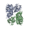 2iobS S: Starting model for refinement C: citing same article ( |
|---|---|
| Similar structure data |
-Links
 PDBj
PDBj


- Assembly
| Deposited unit | 
| ||||||||
|---|---|---|---|---|---|---|---|---|---|
| 1 |
| ||||||||
| Unit cell |
|
-Components
| #1: Protein | Mass: 46025.578 Da / Num. of mol.: 1 Source method: isolated from a genetically manipulated source Source: (gene. exp.) References: UniProt: P33222, Ligases; Forming carbon-nitrogen bonds; Acid-ammonia (or amine) ligases (amide synthases) | ||||
|---|---|---|---|---|---|
| #2: Chemical | ChemComp-SO4 /   Mass: 96.063 Da / Num. of mol.: 6 / Source method: obtained synthetically / Formula: SO4 Mass: 96.063 Da / Num. of mol.: 6 / Source method: obtained synthetically / Formula: SO4#3: Water | ChemComp-HOH / |  Mass: 18.015 Da / Num. of mol.: 289 / Source method: isolated from a natural source / Formula: H2O Mass: 18.015 Da / Num. of mol.: 289 / Source method: isolated from a natural source / Formula: H2OHas ligand of interest | N | |
-Experimental details
-Experiment
| Experiment | Method: X-RAY DIFFRACTION / Number of used crystals: 1 |
|---|
- Sample preparation
| Crystal | Density Matthews: 2.86 Å3/Da / Density % sol: 56.93 % |
|---|---|
| Crystal grow | Temperature: 289.15 K / Method: vapor diffusion, sitting drop Details: 200 mM lithium sulfate monohydrate, 0.1 M Tris-HCl pH 8.5, 20% PEG 4000 |
-Data collection
| Diffraction | Mean temperature: 100 K / Serial crystal experiment: N | ||||||||||||||||||||||||||||||
|---|---|---|---|---|---|---|---|---|---|---|---|---|---|---|---|---|---|---|---|---|---|---|---|---|---|---|---|---|---|---|---|
| Diffraction source | Source: SYNCHROTRON / Site: Australian Synchrotron  / Beamline: MX2 / Wavelength: 0.9537 Å / Beamline: MX2 / Wavelength: 0.9537 Å | ||||||||||||||||||||||||||||||
| Detector | Type: DECTRIS EIGER X 16M / Detector: PIXEL / Date: Jul 7, 2020 | ||||||||||||||||||||||||||||||
| Radiation | Protocol: SINGLE WAVELENGTH / Monochromatic (M) / Laue (L): M / Scattering type: x-ray | ||||||||||||||||||||||||||||||
| Radiation wavelength | Wavelength: 0.9537 Å / Relative weight: 1 | ||||||||||||||||||||||||||||||
| Reflection | Resolution: 1.9→44.42 Å / Num. obs: 42317 / % possible obs: 100 % / Redundancy: 13.2 % / CC1/2: 0.999 / Rmerge(I) obs: 0.088 / Rpim(I) all: 0.025 / Rrim(I) all: 0.092 / Net I/σ(I): 15.5 / Num. measured all: 559446 / Scaling rejects: 19 | ||||||||||||||||||||||||||||||
| Reflection shell | Diffraction-ID: 1
|
- Processing
| Software |
| ||||||||||||||||||||||||||||||||||||||||||||||||||||||||||||||||||||||||||||||||||||||||||||||||
|---|---|---|---|---|---|---|---|---|---|---|---|---|---|---|---|---|---|---|---|---|---|---|---|---|---|---|---|---|---|---|---|---|---|---|---|---|---|---|---|---|---|---|---|---|---|---|---|---|---|---|---|---|---|---|---|---|---|---|---|---|---|---|---|---|---|---|---|---|---|---|---|---|---|---|---|---|---|---|---|---|---|---|---|---|---|---|---|---|---|---|---|---|---|---|---|---|---|
| Refinement | Method to determine structure: MOLECULAR REPLACEMENT Starting model: 2IOB Resolution: 1.9→36.62 Å / SU ML: 0.2 / Cross valid method: THROUGHOUT / σ(F): 1.33 / Phase error: 22.5 / Stereochemistry target values: ML
| ||||||||||||||||||||||||||||||||||||||||||||||||||||||||||||||||||||||||||||||||||||||||||||||||
| Solvent computation | Shrinkage radii: 0.9 Å / VDW probe radii: 1.11 Å / Solvent model: FLAT BULK SOLVENT MODEL | ||||||||||||||||||||||||||||||||||||||||||||||||||||||||||||||||||||||||||||||||||||||||||||||||
| Displacement parameters | Biso max: 94.11 Å2 / Biso mean: 40.7611 Å2 / Biso min: 24.29 Å2 | ||||||||||||||||||||||||||||||||||||||||||||||||||||||||||||||||||||||||||||||||||||||||||||||||
| Refinement step | Cycle: final / Resolution: 1.9→36.62 Å
| ||||||||||||||||||||||||||||||||||||||||||||||||||||||||||||||||||||||||||||||||||||||||||||||||
| LS refinement shell | Refine-ID: X-RAY DIFFRACTION / Rfactor Rfree error: 0 / Total num. of bins used: 15 / % reflection obs: 100 %
|
