 Movie
Movie Controller

[English] 日本語
 Yorodumi
- PDB-7ui3: Apo-form of Human Tryptophan 2,3-Dioxygenase Induced by NADH Binding -
+ Open data

- Basic information
| Entry | Database: PDB / ID: 7ui3 | |||||||||
|---|---|---|---|---|---|---|---|---|---|---|
| Title | Apo-form of Human Tryptophan 2,3-Dioxygenase Induced by NADH Binding | |||||||||
 Components Components | Tryptophan 2,3-dioxygenase | |||||||||
 Keywords Keywords | OXIDOREDUCTASE / Tryptophan 2 / 3-Dioxygenase / apo-form / NADH / OXYGEN BINDING | |||||||||
| Function / homology |  Function and homology information Function and homology informationresponse to nitroglycerin / tryptophan 2,3-dioxygenase / : / L-tryptophan 2,3-dioxygenase activity / : / response to cortisol / Tryptophan catabolism / amino acid binding / oxygen binding / protein homotetramerization ...response to nitroglycerin / tryptophan 2,3-dioxygenase / : / L-tryptophan 2,3-dioxygenase activity / : / response to cortisol / Tryptophan catabolism / amino acid binding / oxygen binding / protein homotetramerization / response to ethanol / heme binding / metal ion binding / identical protein binding / cytosol Similarity search - Function | |||||||||
| Biological species |  Homo sapiens (human) Homo sapiens (human) | |||||||||
| Method |  X-RAY DIFFRACTION / SYNCHROTRON / MOLECULAR REPLACEMENT / Resolution: 3.18 Å X-RAY DIFFRACTION / SYNCHROTRON / MOLECULAR REPLACEMENT / Resolution: 3.18 Å | |||||||||
 Authors Authors | Yeh, S.-R. / Geeraerts, Z. | |||||||||
| Funding support |  United States, 2items United States, 2items
| |||||||||
 Citation Citation | Journal: To Be Published Title: Apo-form of Human Tryptophan 2,3-Dioxygenase Induced by NADH Binding Authors: Yeh, S.-R. / Geeraerts, Z. | |||||||||
| History |
|
- Structure visualization
| Structure viewer | Molecule: MolmilJmol/JSmol |
|---|
- Downloads & links
-Download
| PDBx/mmCIF format | 7ui3.cif.gz | 448.1 KB | Display | PDBx/mmCIF format |
|---|---|---|---|---|
| PDB format | pdb7ui3.ent.gz | 355.1 KB | Display | PDB format |
| PDBx/mmJSON format | 7ui3.json.gz | Tree view | PDBx/mmJSON format | |
| Others |  Other downloads Other downloads |
-Validation report
| Arichive directory | https://data.pdbj.org/pub/pdb/validation_reports/ui/7ui3ftp://data.pdbj.org/pub/pdb/validation_reports/ui/7ui3 | HTTPS FTP |
|---|
-Related structure data
| Related structure data | 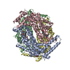 6pyzS S: Starting model for refinement |
|---|---|
| Similar structure data |
-Links
 PDBj
PDBj- Assembly
| Deposited unit | 
| ||||||||
|---|---|---|---|---|---|---|---|---|---|
| 1 |
| ||||||||
| Unit cell |
|
-Components
| #1: Protein | Mass: 45182.535 Da / Num. of mol.: 4 / Fragment: UNP residues 18-389 Source method: isolated from a genetically manipulated source Source: (gene. exp.) Homo sapiens (human) / Gene: TDO2, TDO / Production host:  #2: Chemical | ChemComp-ZIQ /   Type: L-peptide linking / Mass: 218.252 Da / Num. of mol.: 4 / Source method: obtained synthetically / Formula: C12H14N2O2 Type: L-peptide linking / Mass: 218.252 Da / Num. of mol.: 4 / Source method: obtained synthetically / Formula: C12H14N2O2Has ligand of interest | N | Has protein modification | N | |
|---|
-Experimental details
-Experiment
| Experiment | Method: X-RAY DIFFRACTION / Number of used crystals: 1 |
|---|
- Sample preparation
| Crystal | Density Matthews: 2.77 Å3/Da / Density % sol: 55.55 % |
|---|---|
| Crystal grow | Temperature: 293 K / Method: microbatch / pH: 5.6 Details: 50 mM Sodium Citrate, pH 5.6 2% Tacsimate PEG 3350 (4-12%) |
-Data collection
| Diffraction | Mean temperature: 100 K / Serial crystal experiment: N | |||||||||||||||||||||
|---|---|---|---|---|---|---|---|---|---|---|---|---|---|---|---|---|---|---|---|---|---|---|
| Diffraction source | Source: SYNCHROTRON / Site: SSRL / Beamline: BL9-2 / Wavelength: 1.54975 Å | |||||||||||||||||||||
| Detector | Type: DECTRIS PILATUS 6M / Detector: PIXEL / Date: Nov 21, 2021 Details: Rh coated flat bent M0, toroidal focusing post-monochromator M1 | |||||||||||||||||||||
| Radiation | Monochromator: Si(111) and Si(220) double crystal / Protocol: SINGLE WAVELENGTH / Monochromatic (M) / Laue (L): M / Scattering type: x-ray | |||||||||||||||||||||
| Radiation wavelength | Wavelength: 1.54975 Å / Relative weight: 1 | |||||||||||||||||||||
| Reflection | Resolution: 3.18→19.974 Å / Num. obs: 34269 / % possible obs: 99.5 % / Redundancy: 12.9 % / CC1/2: 1 / Rmerge(I) obs: 0.098 / Rpim(I) all: 0.04 / Rrim(I) all: 0.106 / Net I/σ(I): 18.5 | |||||||||||||||||||||
| Reflection shell | Diffraction-ID: 1
|
- Processing
| Software |
| |||||||||||||||||||||||||||||||||||||||||||||||||||||||||||||||||||||||||||||||||||||||||||||||||||||||||||||||||||||||||||||||||||||||||||||||||||||||||||||||||||||||||||||||
|---|---|---|---|---|---|---|---|---|---|---|---|---|---|---|---|---|---|---|---|---|---|---|---|---|---|---|---|---|---|---|---|---|---|---|---|---|---|---|---|---|---|---|---|---|---|---|---|---|---|---|---|---|---|---|---|---|---|---|---|---|---|---|---|---|---|---|---|---|---|---|---|---|---|---|---|---|---|---|---|---|---|---|---|---|---|---|---|---|---|---|---|---|---|---|---|---|---|---|---|---|---|---|---|---|---|---|---|---|---|---|---|---|---|---|---|---|---|---|---|---|---|---|---|---|---|---|---|---|---|---|---|---|---|---|---|---|---|---|---|---|---|---|---|---|---|---|---|---|---|---|---|---|---|---|---|---|---|---|---|---|---|---|---|---|---|---|---|---|---|---|---|---|---|---|---|---|
| Refinement | Method to determine structure: MOLECULAR REPLACEMENT Starting model: 6PYZ Resolution: 3.18→19.974 Å / Cor.coef. Fo:Fc: 0.948 / Cor.coef. Fo:Fc free: 0.934 / WRfactor Rfree: 0.26 / WRfactor Rwork: 0.227 / SU B: 28.651 / SU ML: 0.452 / Average fsc free: 0.8269 / Average fsc work: 0.8441 / Cross valid method: FREE R-VALUE / ESU R Free: 0.479 Details: Hydrogens have been added in their riding positions
| |||||||||||||||||||||||||||||||||||||||||||||||||||||||||||||||||||||||||||||||||||||||||||||||||||||||||||||||||||||||||||||||||||||||||||||||||||||||||||||||||||||||||||||||
| Solvent computation | Ion probe radii: 0.9 Å / Shrinkage radii: 0.9 Å / VDW probe radii: 1 Å / Solvent model: MASK BULK SOLVENT | |||||||||||||||||||||||||||||||||||||||||||||||||||||||||||||||||||||||||||||||||||||||||||||||||||||||||||||||||||||||||||||||||||||||||||||||||||||||||||||||||||||||||||||||
| Displacement parameters | Biso mean: 106.154 Å2
| |||||||||||||||||||||||||||||||||||||||||||||||||||||||||||||||||||||||||||||||||||||||||||||||||||||||||||||||||||||||||||||||||||||||||||||||||||||||||||||||||||||||||||||||
| Refinement step | Cycle: LAST / Resolution: 3.18→19.974 Å
| |||||||||||||||||||||||||||||||||||||||||||||||||||||||||||||||||||||||||||||||||||||||||||||||||||||||||||||||||||||||||||||||||||||||||||||||||||||||||||||||||||||||||||||||
| Refine LS restraints |
| |||||||||||||||||||||||||||||||||||||||||||||||||||||||||||||||||||||||||||||||||||||||||||||||||||||||||||||||||||||||||||||||||||||||||||||||||||||||||||||||||||||||||||||||
| LS refinement shell |
|
