 Movie
Movie Controller

[English] 日本語
 Yorodumi
- PDB-7ui2: The crystal structure of 15kDa Phlebotomus papatasi salivary prot... -
+ Open data

- Basic information
| Entry | Database: PDB / ID: 7ui2 | ||||||
|---|---|---|---|---|---|---|---|
| Title | The crystal structure of 15kDa Phlebotomus papatasi salivary protein Ppsp15. | ||||||
 Components Components | SP15 protein | ||||||
 Keywords Keywords | PROTEIN BINDING / SALIVARY / SANDFLY / VACCINE / LEISHMANIA | ||||||
| Function / homology | Pheromone/general odorant binding protein superfamily / odorant binding / extracellular region / ACETATE ION / SP15 protein Function and homology information Function and homology information | ||||||
| Biological species |  Phlebotomus papatasi (insect) Phlebotomus papatasi (insect) | ||||||
| Method |  X-RAY DIFFRACTION / SYNCHROTRON / MOLECULAR REPLACEMENT / Resolution: 1.19 Å X-RAY DIFFRACTION / SYNCHROTRON / MOLECULAR REPLACEMENT / Resolution: 1.19 Å | ||||||
 Authors Authors | Tolbert, W.D. / Pazgier, M. | ||||||
| Funding support | 1items
| ||||||
 Citation Citation | Journal: To Be Published Title: The crystal structure of 15kDa Phlebotomus papatasi salivary protein Ppsp15. Authors: Tolbert, W.D. / Pazgier, M. | ||||||
| History |
|
- Structure visualization
| Structure viewer | Molecule: MolmilJmol/JSmol |
|---|
- Downloads & links
-Download
| PDBx/mmCIF format | 7ui2.cif.gz | 125.1 KB | Display | PDBx/mmCIF format |
|---|---|---|---|---|
| PDB format | pdb7ui2.ent.gz | 96 KB | Display | PDB format |
| PDBx/mmJSON format | 7ui2.json.gz | Tree view | PDBx/mmJSON format | |
| Others |  Other downloads Other downloads |
-Validation report
| Summary document | 7ui2_validation.pdf.gz | 451.3 KB | Display | wwPDB validaton report |
|---|---|---|---|---|
| Full document | 7ui2_full_validation.pdf.gz | 453.9 KB | Display | |
| Data in XML | 7ui2_validation.xml.gz | 15.6 KB | Display | |
| Data in CIF | 7ui2_validation.cif.gz | 23 KB | Display | |
| Arichive directory | https://data.pdbj.org/pub/pdb/validation_reports/ui/7ui2ftp://data.pdbj.org/pub/pdb/validation_reports/ui/7ui2 | HTTPS FTP |
-Related structure data
| Related structure data | 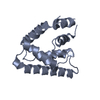 4ozdS S: Starting model for refinement |
|---|---|
| Similar structure data |
-Links
 PDBj
PDBj
- Assembly
| Deposited unit | 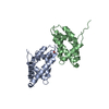
| ||||||||
|---|---|---|---|---|---|---|---|---|---|
| 1 | 
| ||||||||
| 2 | 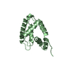
| ||||||||
| Unit cell |
|
-Components
| #1: Protein | Mass: 15174.511 Da / Num. of mol.: 2 Source method: isolated from a genetically manipulated source Source: (gene. exp.) Phlebotomus papatasi (insect) / Production host:  Komagataella pastoris (fungus) / References: UniProt: F8R290 Komagataella pastoris (fungus) / References: UniProt: F8R290#2: Chemical | ChemComp-ACT / |   Mass: 59.044 Da / Num. of mol.: 1 / Source method: obtained synthetically / Formula: C2H3O2 Mass: 59.044 Da / Num. of mol.: 1 / Source method: obtained synthetically / Formula: C2H3O2#3: Water | ChemComp-HOH / |  Mass: 18.015 Da / Num. of mol.: 351 / Source method: isolated from a natural source / Formula: H2O Mass: 18.015 Da / Num. of mol.: 351 / Source method: isolated from a natural source / Formula: H2OHas ligand of interest | N | Has protein modification | Y | |
|---|
-Experimental details
-Experiment
| Experiment | Method: X-RAY DIFFRACTION / Number of used crystals: 1 |
|---|
- Sample preparation
| Crystal | Density Matthews: 1.91 Å3/Da / Density % sol: 35.47 % |
|---|---|
| Crystal grow | Temperature: 294 K / Method: vapor diffusion, hanging drop / pH: 8.5 Details: 25% PEG 3350 0.2 M sodium chloride 0.1 M Tris-HCl pH 8.5 |
-Data collection
| Diffraction | Mean temperature: 100 K / Serial crystal experiment: N |
|---|---|
| Diffraction source | Source: SYNCHROTRON / Site: SSRL  / Beamline: BL12-2 / Wavelength: 0.97946 Å / Beamline: BL12-2 / Wavelength: 0.97946 Å |
| Detector | Type: DECTRIS PILATUS 6M / Detector: PIXEL / Date: Mar 18, 2022 |
| Radiation | Monochromator: Si(111) / Protocol: SINGLE WAVELENGTH / Monochromatic (M) / Laue (L): M / Scattering type: x-ray |
| Radiation wavelength | Wavelength: 0.97946 Å / Relative weight: 1 |
| Reflection | Resolution: 1.19→50 Å / Num. obs: 128818 / % possible obs: 96.2 % / Redundancy: 3.3 % / CC1/2: 0.976 / Rpim(I) all: 0.066 / Rsym value: 0.103 / Net I/σ(I): 2.4 |
| Reflection shell | Resolution: 1.19→1.26 Å / Mean I/σ(I) obs: 0.6 / Num. unique obs: 9811 / CC1/2: 0.423 / Rpim(I) all: 0.484 / Rsym value: 0.75 / % possible all: 95.6 |
- Processing
| Software |
| |||||||||||||||||||||||||||||||||||||||||||||||||||||||||||||||||||||||||||||||||||||||||||||||||||||||||||||||||||||||||||||||||||||||||||||||||||
|---|---|---|---|---|---|---|---|---|---|---|---|---|---|---|---|---|---|---|---|---|---|---|---|---|---|---|---|---|---|---|---|---|---|---|---|---|---|---|---|---|---|---|---|---|---|---|---|---|---|---|---|---|---|---|---|---|---|---|---|---|---|---|---|---|---|---|---|---|---|---|---|---|---|---|---|---|---|---|---|---|---|---|---|---|---|---|---|---|---|---|---|---|---|---|---|---|---|---|---|---|---|---|---|---|---|---|---|---|---|---|---|---|---|---|---|---|---|---|---|---|---|---|---|---|---|---|---|---|---|---|---|---|---|---|---|---|---|---|---|---|---|---|---|---|---|---|---|---|
| Refinement | Method to determine structure: MOLECULAR REPLACEMENT Starting model: 4OZD Resolution: 1.19→40.87 Å / Cross valid method: FREE R-VALUE / σ(F): 0 / Phase error: 23.49 / Stereochemistry target values: TWIN_LSQ_F
| |||||||||||||||||||||||||||||||||||||||||||||||||||||||||||||||||||||||||||||||||||||||||||||||||||||||||||||||||||||||||||||||||||||||||||||||||||
| Solvent computation | Shrinkage radii: 0.9 Å / VDW probe radii: 1.11 Å / Solvent model: FLAT BULK SOLVENT MODEL | |||||||||||||||||||||||||||||||||||||||||||||||||||||||||||||||||||||||||||||||||||||||||||||||||||||||||||||||||||||||||||||||||||||||||||||||||||
| Refinement step | Cycle: LAST / Resolution: 1.19→40.87 Å
| |||||||||||||||||||||||||||||||||||||||||||||||||||||||||||||||||||||||||||||||||||||||||||||||||||||||||||||||||||||||||||||||||||||||||||||||||||
| Refine LS restraints |
| |||||||||||||||||||||||||||||||||||||||||||||||||||||||||||||||||||||||||||||||||||||||||||||||||||||||||||||||||||||||||||||||||||||||||||||||||||
| LS refinement shell |
| |||||||||||||||||||||||||||||||||||||||||||||||||||||||||||||||||||||||||||||||||||||||||||||||||||||||||||||||||||||||||||||||||||||||||||||||||||
| Refinement TLS params. | Method: refined / Refine-ID: X-RAY DIFFRACTION
| |||||||||||||||||||||||||||||||||||||||||||||||||||||||||||||||||||||||||||||||||||||||||||||||||||||||||||||||||||||||||||||||||||||||||||||||||||
| Refinement TLS group |
|
