 Movie
Movie Controller

[English] 日本語
 Yorodumi
- PDB-7ug8: Crystal structure of a solute receptor from Synechococcus CC9311 ... -
+ Open data

- Basic information
| Entry | Database: PDB / ID: 7ug8 | |||||||||
|---|---|---|---|---|---|---|---|---|---|---|
| Title | Crystal structure of a solute receptor from Synechococcus CC9311 in complex with alpha-ketovaleric and calcium | |||||||||
 Components Components | Ketoacid-binding protein | |||||||||
 Keywords Keywords | TRANSPORT PROTEIN / Substrate-binding protein / marine cyanobacteria / ketoacid-binding protein | |||||||||
| Function / homology |  Function and homology information Function and homology informationorganic acid transport / tripartite ATP-independent periplasmic transporter complex / organic acid binding / transmembrane transport / metal ion binding Similarity search - Function | |||||||||
| Biological species |  Synechococcus sp. (bacteria) Synechococcus sp. (bacteria) | |||||||||
| Method |  X-RAY DIFFRACTION / SYNCHROTRON / SAD / Resolution: 1.796 Å X-RAY DIFFRACTION / SYNCHROTRON / SAD / Resolution: 1.796 Å | |||||||||
 Authors Authors | Shah, B.S. / Mikolajek, H. / Orr, C.M. / Mykhaylyk, V. / Owens, R.J. / Paulsen, I.T. | |||||||||
| Funding support |  Australia, 2items Australia, 2items
| |||||||||
 Citation Citation | Journal: To Be Published Title: Crystal structure of a solute receptor from Synechococcus CC9311 in complex with alpha-ketovaleric and calcium Authors: Shah, B.S. / Mikolajek, H. / Orr, C.M. / Mykhaylyk, V. / Owens, R. / Paulsen, I.T. | |||||||||
| History |
|
- Structure visualization
| Structure viewer | Molecule: MolmilJmol/JSmol |
|---|
- Downloads & links
-Download
| PDBx/mmCIF format | 7ug8.cif.gz | 264.2 KB | Display | PDBx/mmCIF format |
|---|---|---|---|---|
| PDB format | pdb7ug8.ent.gz | 209.7 KB | Display | PDB format |
| PDBx/mmJSON format | 7ug8.json.gz | Tree view | PDBx/mmJSON format | |
| Others |  Other downloads Other downloads |
-Validation report
| Arichive directory | https://data.pdbj.org/pub/pdb/validation_reports/ug/7ug8ftp://data.pdbj.org/pub/pdb/validation_reports/ug/7ug8 | HTTPS FTP |
|---|
-Related structure data
| Similar structure data |
|---|
-Links
 PDBj
PDBj


- Assembly
| Deposited unit | 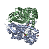
| ||||||||||||||||||
|---|---|---|---|---|---|---|---|---|---|---|---|---|---|---|---|---|---|---|---|
| 1 |
| ||||||||||||||||||
| Unit cell |
| ||||||||||||||||||
| Noncrystallographic symmetry (NCS) | NCS domain:
NCS domain segments: Component-ID: 1 / Ens-ID: 1 / Beg auth comp-ID: GLY / Beg label comp-ID: GLY / End auth comp-ID: PHE / End label comp-ID: PHE / Auth seq-ID: 7 - 334 / Label seq-ID: 7 - 334
NCS ensembles : (Details: Local NCS retraints between domains: 1 2) |
-Components
| #1: Protein | Mass: 38460.270 Da / Num. of mol.: 2 Source method: isolated from a genetically manipulated source Source: (gene. exp.) Synechococcus sp. (strain CC9311) (bacteria)Strain: CC9311 / Gene: sync_2776 / Production host: #2: Chemical | 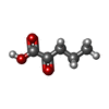  Mass: 116.115 Da / Num. of mol.: 2 / Source method: obtained synthetically / Formula: C5H8O3 / Feature type: SUBJECT OF INVESTIGATION Mass: 116.115 Da / Num. of mol.: 2 / Source method: obtained synthetically / Formula: C5H8O3 / Feature type: SUBJECT OF INVESTIGATION#3: Chemical |   Mass: 62.068 Da / Num. of mol.: 2 / Source method: obtained synthetically / Formula: C2H6O2 Mass: 62.068 Da / Num. of mol.: 2 / Source method: obtained synthetically / Formula: C2H6O2#4: Chemical |   Mass: 40.078 Da / Num. of mol.: 2 / Source method: obtained synthetically / Formula: Ca Mass: 40.078 Da / Num. of mol.: 2 / Source method: obtained synthetically / Formula: Ca#5: Water | ChemComp-HOH / |  Mass: 18.015 Da / Num. of mol.: 255 / Source method: isolated from a natural source / Formula: H2O Mass: 18.015 Da / Num. of mol.: 255 / Source method: isolated from a natural source / Formula: H2OHas ligand of interest | Y | |
|---|
-Experimental details
-Experiment
| Experiment | Method: X-RAY DIFFRACTION / Number of used crystals: 1 |
|---|
- Sample preparation
| Crystal | Density Matthews: 2.05 Å3/Da / Density % sol: 39.99 % |
|---|---|
| Crystal grow | Temperature: 295 K / Method: vapor diffusion, sitting drop / pH: 7.5 Details: 0.02 M Sodium/Potassium Phosphate, 0.1M Bis Tris propane 7.5, 20% Peg 3350 Cryo: 30 % Peg 400 |
-Data collection
| Diffraction | Mean temperature: 50 K / Serial crystal experiment: N | ||||||||||||||||||||||||||||||||||||||||||||||||||
|---|---|---|---|---|---|---|---|---|---|---|---|---|---|---|---|---|---|---|---|---|---|---|---|---|---|---|---|---|---|---|---|---|---|---|---|---|---|---|---|---|---|---|---|---|---|---|---|---|---|---|---|
| Diffraction source | Source: SYNCHROTRON / Site: Diamond  / Beamline: I23 / Wavelength: 2.7552, 3.1790, 3.4925, 4.7686 / Beamline: I23 / Wavelength: 2.7552, 3.1790, 3.4925, 4.7686 | ||||||||||||||||||||||||||||||||||||||||||||||||||
| Detector | Type: DECTRIS PILATUS 12M / Detector: PIXEL / Date: Dec 12, 2019 | ||||||||||||||||||||||||||||||||||||||||||||||||||
| Radiation | Protocol: SINGLE WAVELENGTH / Monochromatic (M) / Laue (L): M / Scattering type: x-ray | ||||||||||||||||||||||||||||||||||||||||||||||||||
| Radiation wavelength |
| ||||||||||||||||||||||||||||||||||||||||||||||||||
| Reflection | Entry-ID: 7UG8
| ||||||||||||||||||||||||||||||||||||||||||||||||||
| Reflection shell | Resolution: 9.06→50.23 Å / Redundancy: 23.3 % / Rmerge(I) obs: 0.039 / Num. unique obs: 451 / CC1/2: 0.999 / Rpim(I) all: 0.012 / Rrim(I) all: 0.041 |
- Processing
| Software |
| |||||||||||||||||||||||||||||||||||||||||||||||||||||||||||||||||||||||||||||||||||||||||||||||||||||||||||||||||||||||||||||||||||||||||||||||||||||||||||||||||||||
|---|---|---|---|---|---|---|---|---|---|---|---|---|---|---|---|---|---|---|---|---|---|---|---|---|---|---|---|---|---|---|---|---|---|---|---|---|---|---|---|---|---|---|---|---|---|---|---|---|---|---|---|---|---|---|---|---|---|---|---|---|---|---|---|---|---|---|---|---|---|---|---|---|---|---|---|---|---|---|---|---|---|---|---|---|---|---|---|---|---|---|---|---|---|---|---|---|---|---|---|---|---|---|---|---|---|---|---|---|---|---|---|---|---|---|---|---|---|---|---|---|---|---|---|---|---|---|---|---|---|---|---|---|---|---|---|---|---|---|---|---|---|---|---|---|---|---|---|---|---|---|---|---|---|---|---|---|---|---|---|---|---|---|---|---|---|---|
| Refinement | Method to determine structure: SAD / Resolution: 1.796→50.277 Å / Cor.coef. Fo:Fc: 0.977 / Cor.coef. Fo:Fc free: 0.972 / SU B: 3.728 / SU ML: 0.103 / Cross valid method: FREE R-VALUE / ESU R: 0.146 / ESU R Free: 0.121 Details: Hydrogens have been added in their riding positions
| |||||||||||||||||||||||||||||||||||||||||||||||||||||||||||||||||||||||||||||||||||||||||||||||||||||||||||||||||||||||||||||||||||||||||||||||||||||||||||||||||||||
| Solvent computation | Ion probe radii: 0.8 Å / Shrinkage radii: 0.8 Å / VDW probe radii: 1.2 Å / Solvent model: MASK BULK SOLVENT | |||||||||||||||||||||||||||||||||||||||||||||||||||||||||||||||||||||||||||||||||||||||||||||||||||||||||||||||||||||||||||||||||||||||||||||||||||||||||||||||||||||
| Displacement parameters | Biso mean: 44.394 Å2
| |||||||||||||||||||||||||||||||||||||||||||||||||||||||||||||||||||||||||||||||||||||||||||||||||||||||||||||||||||||||||||||||||||||||||||||||||||||||||||||||||||||
| Refinement step | Cycle: LAST / Resolution: 1.796→50.277 Å
| |||||||||||||||||||||||||||||||||||||||||||||||||||||||||||||||||||||||||||||||||||||||||||||||||||||||||||||||||||||||||||||||||||||||||||||||||||||||||||||||||||||
| Refine LS restraints |
| |||||||||||||||||||||||||||||||||||||||||||||||||||||||||||||||||||||||||||||||||||||||||||||||||||||||||||||||||||||||||||||||||||||||||||||||||||||||||||||||||||||
| Refine LS restraints NCS |
| |||||||||||||||||||||||||||||||||||||||||||||||||||||||||||||||||||||||||||||||||||||||||||||||||||||||||||||||||||||||||||||||||||||||||||||||||||||||||||||||||||||
| LS refinement shell |
|
