 Movie
Movie Controller


+ Open data

- Basic information
| Entry | Database: PDB / ID: 7ufo | ||||||
|---|---|---|---|---|---|---|---|
| Title | Structure of PfCSP peptide 21 with antibody P3-42 | ||||||
 Components Components |
| ||||||
 Keywords Keywords | IMMUNE SYSTEM / Malaria / Circumsporozoite | ||||||
| Function / homology |  Function and homology information Function and homology information | ||||||
| Biological species |  Homo sapiens (human) Homo sapiens (human) | ||||||
| Method |  X-RAY DIFFRACTION / SYNCHROTRON / MOLECULAR REPLACEMENT / Resolution: 1.8 Å X-RAY DIFFRACTION / SYNCHROTRON / MOLECULAR REPLACEMENT / Resolution: 1.8 Å | ||||||
 Authors Authors | Tripathi, P. / Kwong, P.D. | ||||||
| Funding support |  United States, 1items United States, 1items
| ||||||
 Citation Citation | Journal: To Be Published Title: Structure of PfCSP peptide 21 with antibody P3-42 Authors: Tripathi, P. / Kwong, P.D. | ||||||
| History |
|
- Structure visualization
| Structure viewer | Molecule: MolmilJmol/JSmol |
|---|
- Downloads & links
-Download
| PDBx/mmCIF format | 7ufo.cif.gz | 108.3 KB | Display | PDBx/mmCIF format |
|---|---|---|---|---|
| PDB format | pdb7ufo.ent.gz | 80.3 KB | Display | PDB format |
| PDBx/mmJSON format | 7ufo.json.gz | Tree view | PDBx/mmJSON format | |
| Others |  Other downloads Other downloads |
-Validation report
| Summary document | 7ufo_validation.pdf.gz | 465.2 KB | Display | wwPDB validaton report |
|---|---|---|---|---|
| Full document | 7ufo_full_validation.pdf.gz | 467.7 KB | Display | |
| Data in XML | 7ufo_validation.xml.gz | 21.1 KB | Display | |
| Data in CIF | 7ufo_validation.cif.gz | 30.6 KB | Display | |
| Arichive directory | https://data.pdbj.org/pub/pdb/validation_reports/uf/7ufoftp://data.pdbj.org/pub/pdb/validation_reports/uf/7ufo | HTTPS FTP |
-Related structure data
| Related structure data | 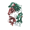 6b5mS S: Starting model for refinement |
|---|---|
| Similar structure data |
-Links
 PDBj
PDBj

- Assembly
| Deposited unit | 
| |||||||||
|---|---|---|---|---|---|---|---|---|---|---|
| 1 |
| |||||||||
| Unit cell |
| |||||||||
| Components on special symmetry positions |
|
-Components
-Protein/peptide , 1 types, 1 molecules A
| #3: Protein/peptide | Mass: 1562.553 Da / Num. of mol.: 1 / Source method: obtained synthetically Source: (synth.) References: UniProt: P08307 |
|---|
-Antibody , 2 types, 2 molecules HL
| #1: Antibody | Mass: 24244.283 Da / Num. of mol.: 1 Source method: isolated from a genetically manipulated source Source: (gene. exp.) Homo sapiens (human) / Production host: Homo sapiens (human) |
|---|---|
| #2: Antibody | Mass: 24229.838 Da / Num. of mol.: 1 Source method: isolated from a genetically manipulated source Source: (gene. exp.) Homo sapiens (human) / Production host: Homo sapiens (human) |
-Non-polymers , 3 types, 300 molecules 


| #4: Chemical |  Mass: 92.094 Da / Num. of mol.: 2 / Source method: obtained synthetically / Formula: C3H8O3 Mass: 92.094 Da / Num. of mol.: 2 / Source method: obtained synthetically / Formula: C3H8O3#5: Chemical |  Mass: 96.063 Da / Num. of mol.: 2 / Source method: obtained synthetically / Formula: SO4 Mass: 96.063 Da / Num. of mol.: 2 / Source method: obtained synthetically / Formula: SO4#6: Water | ChemComp-HOH / | Mass: 18.015 Da / Num. of mol.: 296 / Source method: isolated from a natural source / Formula: H2O |
|---|
-Details
| Has ligand of interest | N |
|---|---|
| Has protein modification | Y |
-Experimental details
-Experiment
| Experiment | Method: X-RAY DIFFRACTION / Number of used crystals: 1 |
|---|
- Sample preparation
| Crystal | Density Matthews: 2.38 Å3/Da / Density % sol: 48.33 % |
|---|---|
| Crystal grow | Temperature: 298 K / Method: vapor diffusion, sitting drop Details: 0.1 M Tris hydrochloride pH 8.5, 2.8 M ammonium sulfate |
-Data collection
| Diffraction | Mean temperature: 100 K / Serial crystal experiment: N |
|---|---|
| Diffraction source | Source: SYNCHROTRON / Site: APS / Beamline: 22-ID / Wavelength: 1 Å |
| Detector | Type: DECTRIS EIGER X 16M / Detector: PIXEL / Date: Jan 14, 2022 |
| Radiation | Protocol: SINGLE WAVELENGTH / Monochromatic (M) / Laue (L): M / Scattering type: x-ray |
| Radiation wavelength | Wavelength: 1 Å / Relative weight: 1 |
| Reflection | Resolution: 1.8→46.43 Å / Num. obs: 42986 / % possible obs: 98.18 % / Redundancy: 3.5 % / CC1/2: 0.988 / Net I/σ(I): 7.51 |
| Reflection shell | Resolution: 1.8→1.86 Å / Num. unique obs: 4118 / CC1/2: 0.765 |
- Processing
| Software |
| ||||||||||||||||||||||||||||||||||||||||||||||||||||||||||||||||||||||||||||||||||||||||||||||||
|---|---|---|---|---|---|---|---|---|---|---|---|---|---|---|---|---|---|---|---|---|---|---|---|---|---|---|---|---|---|---|---|---|---|---|---|---|---|---|---|---|---|---|---|---|---|---|---|---|---|---|---|---|---|---|---|---|---|---|---|---|---|---|---|---|---|---|---|---|---|---|---|---|---|---|---|---|---|---|---|---|---|---|---|---|---|---|---|---|---|---|---|---|---|---|---|---|---|
| Refinement | Method to determine structure: MOLECULAR REPLACEMENT Starting model: 6b5m Resolution: 1.8→46.43 Å / SU ML: 0.19 / Cross valid method: THROUGHOUT / σ(F): 1.35 / Phase error: 24.07 / Stereochemistry target values: ML
| ||||||||||||||||||||||||||||||||||||||||||||||||||||||||||||||||||||||||||||||||||||||||||||||||
| Solvent computation | Shrinkage radii: 0.9 Å / VDW probe radii: 1.11 Å / Solvent model: FLAT BULK SOLVENT MODEL | ||||||||||||||||||||||||||||||||||||||||||||||||||||||||||||||||||||||||||||||||||||||||||||||||
| Displacement parameters | Biso max: 69.16 Å2 / Biso mean: 21.8196 Å2 / Biso min: 6.37 Å2 | ||||||||||||||||||||||||||||||||||||||||||||||||||||||||||||||||||||||||||||||||||||||||||||||||
| Refinement step | Cycle: final / Resolution: 1.8→46.43 Å
| ||||||||||||||||||||||||||||||||||||||||||||||||||||||||||||||||||||||||||||||||||||||||||||||||
| LS refinement shell | Refine-ID: X-RAY DIFFRACTION / Rfactor Rfree error: 0
|
