 Movie
Movie Controller

[English] 日本語
 Yorodumi
- PDB-7ud1: The Crystal Structure of Apo Monomer F57:H:H:H:R58 HCRBPII with H... -
+ Open data

- Basic information
| Entry | Database: PDB / ID: 7ud1 | ||||||
|---|---|---|---|---|---|---|---|
| Title | The Crystal Structure of Apo Monomer F57:H:H:H:R58 HCRBPII with Histidine Insertion in the Hinge Loop Region at 1.3 Angstrom Resolution | ||||||
 Components Components | Retinol-binding protein 2 | ||||||
 Keywords Keywords | LIPID BINDING PROTEIN / LBP / CRBPII | ||||||
| Function / homology |  Function and homology information Function and homology informationvitamin A metabolic process / retinoid binding / retinal binding / molecular carrier activity / retinol binding / epidermis development / fatty acid transport / Retinoid metabolism and transport / fatty acid binding / nucleus / cytosol Similarity search - Function | ||||||
| Biological species |  Homo sapiens (human) Homo sapiens (human) | ||||||
| Method |  X-RAY DIFFRACTION / SYNCHROTRON / MOLECULAR REPLACEMENT / Resolution: 1.32 Å X-RAY DIFFRACTION / SYNCHROTRON / MOLECULAR REPLACEMENT / Resolution: 1.32 Å | ||||||
 Authors Authors | Ghanbarpour, A. / Geiger, J. | ||||||
| Funding support |  United States, 1items United States, 1items
| ||||||
 Citation Citation | Journal: To Be Published Title: The Crystal Structure of Apo Monomer F57:H:H:H:R58 HCRBPII with Histidine Insertion in the Hinge Loop Region at 1.3 Angstrom Resolution Authors: Ghanbarpour, A. / Geiger, J. | ||||||
| History |
|
- Structure visualization
| Structure viewer | Molecule: MolmilJmol/JSmol |
|---|
- Downloads & links
-Download
| PDBx/mmCIF format | 7ud1.cif.gz | 160.9 KB | Display | PDBx/mmCIF format |
|---|---|---|---|---|
| PDB format | pdb7ud1.ent.gz | 104.9 KB | Display | PDB format |
| PDBx/mmJSON format | 7ud1.json.gz | Tree view | PDBx/mmJSON format | |
| Others |  Other downloads Other downloads |
-Validation report
| Arichive directory | https://data.pdbj.org/pub/pdb/validation_reports/ud/7ud1ftp://data.pdbj.org/pub/pdb/validation_reports/ud/7ud1 | HTTPS FTP |
|---|
-Related structure data
| Related structure data |  2rcqS S: Starting model for refinement |
|---|---|
| Similar structure data |
-Links
 PDBj
PDBj


- Assembly
| Deposited unit | 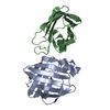
| ||||||||||||
|---|---|---|---|---|---|---|---|---|---|---|---|---|---|
| 1 | 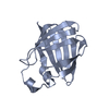
| ||||||||||||
| 2 | 
| ||||||||||||
| Unit cell |
|
-Components
| #1: Protein | Mass: 16011.896 Da / Num. of mol.: 2 Source method: isolated from a genetically manipulated source Source: (gene. exp.) Homo sapiens (human) / Gene: RBP2, CRBP2Production host: Bacterial expression vector pBEN1-SGC (others) References: UniProt: P50120 #2: Water | ChemComp-HOH / |  Mass: 18.015 Da / Num. of mol.: 339 / Source method: isolated from a natural source / Formula: H2O Mass: 18.015 Da / Num. of mol.: 339 / Source method: isolated from a natural source / Formula: H2O |
|---|
-Experimental details
-Experiment
| Experiment | Method: X-RAY DIFFRACTION / Number of used crystals: 1 |
|---|
- Sample preparation
| Crystal | Density Matthews: 1.96 Å3/Da / Density % sol: 37.24 % |
|---|---|
| Crystal grow | Temperature: 298 K / Method: vapor diffusion, hanging drop / Details: PEG 4000, Sodium acetate, ammonium acetate |
-Data collection
| Diffraction | Mean temperature: 100 K / Serial crystal experiment: N |
|---|---|
| Diffraction source | Source: SYNCHROTRON / Site: APS / Beamline: 21-ID-D / Wavelength: 0.97625 Å |
| Detector | Type: DECTRIS EIGER X 9M / Detector: PIXEL / Date: Mar 31, 2018 |
| Radiation | Protocol: SINGLE WAVELENGTH / Monochromatic (M) / Laue (L): M / Scattering type: x-ray |
| Radiation wavelength | Wavelength: 0.97625 Å / Relative weight: 1 |
| Reflection | Resolution: 1.32→29.09 Å / Num. obs: 55379 / % possible obs: 96.6 % / Redundancy: 6.1 % / Biso Wilson estimate: 13 Å2 / Rmerge(I) obs: 0.062 / Rrim(I) all: 0.087 / Net I/σ(I): 27.9 |
| Reflection shell | Resolution: 1.964→2.035 Å / Redundancy: 4.5 % / Rmerge(I) obs: 0.621 / Mean I/σ(I) obs: 2.14 / Num. unique obs: 5345 / Rrim(I) all: 0.602 / % possible all: 93.5 |
- Processing
| Software |
| |||||||||||||||||||||||||||||||||||||||||||||||||||||||||||||||||||||||||||||||||||||||||||||||||||||||||
|---|---|---|---|---|---|---|---|---|---|---|---|---|---|---|---|---|---|---|---|---|---|---|---|---|---|---|---|---|---|---|---|---|---|---|---|---|---|---|---|---|---|---|---|---|---|---|---|---|---|---|---|---|---|---|---|---|---|---|---|---|---|---|---|---|---|---|---|---|---|---|---|---|---|---|---|---|---|---|---|---|---|---|---|---|---|---|---|---|---|---|---|---|---|---|---|---|---|---|---|---|---|---|---|---|---|---|
| Refinement | Method to determine structure: MOLECULAR REPLACEMENT Starting model: 2RCQ Resolution: 1.32→29.09 Å / SU ML: 0.1369 / Cross valid method: FREE R-VALUE / σ(F): 1.34 / Phase error: 20.4963 Stereochemistry target values: GeoStd + Monomer Library + CDL v1.2
| |||||||||||||||||||||||||||||||||||||||||||||||||||||||||||||||||||||||||||||||||||||||||||||||||||||||||
| Solvent computation | Shrinkage radii: 0.9 Å / VDW probe radii: 1.11 Å / Solvent model: FLAT BULK SOLVENT MODEL | |||||||||||||||||||||||||||||||||||||||||||||||||||||||||||||||||||||||||||||||||||||||||||||||||||||||||
| Displacement parameters | Biso mean: 16.73 Å2 | |||||||||||||||||||||||||||||||||||||||||||||||||||||||||||||||||||||||||||||||||||||||||||||||||||||||||
| Refinement step | Cycle: LAST / Resolution: 1.32→29.09 Å
| |||||||||||||||||||||||||||||||||||||||||||||||||||||||||||||||||||||||||||||||||||||||||||||||||||||||||
| Refine LS restraints |
| |||||||||||||||||||||||||||||||||||||||||||||||||||||||||||||||||||||||||||||||||||||||||||||||||||||||||
| LS refinement shell |
| |||||||||||||||||||||||||||||||||||||||||||||||||||||||||||||||||||||||||||||||||||||||||||||||||||||||||
| Refinement TLS params. | Method: refined / Origin x: 60.0223476086 Å / Origin y: -20.5987725934 Å / Origin z: 13.7485355522 Å
| |||||||||||||||||||||||||||||||||||||||||||||||||||||||||||||||||||||||||||||||||||||||||||||||||||||||||
| Refinement TLS group | Selection details: all |
