 Movie
Movie Controller


+ Open data

- Basic information
| Entry | Database: PDB / ID: 7ucr | ||||||
|---|---|---|---|---|---|---|---|
| Title | Joint X-ray/neutron structure of the Sarcin-Ricin loop RNA | ||||||
 Components Components | Sarcin-Ricin loop RNA | ||||||
 Keywords Keywords | RNA / Sarcin-Ricin loop | ||||||
| Function / homology | DEUTERATED WATER / RNA / RNA (> 10) Function and homology information Function and homology information | ||||||
| Biological species |  Homo sapiens (human) Homo sapiens (human) | ||||||
| Method |  X-RAY DIFFRACTION / NEUTRON DIFFRACTION / MOLECULAR REPLACEMENT / Resolution: 1 Å X-RAY DIFFRACTION / NEUTRON DIFFRACTION / MOLECULAR REPLACEMENT / Resolution: 1 Å | ||||||
 Authors Authors | Harp, J.M. / Egli, M.E. / Pallan, P.S. / Coates, L. | ||||||
| Funding support | 1items
| ||||||
 Citation Citation | Journal: Nucleic Acids Res. / Year: 2022 Title: Cryo neutron crystallography demonstrates influence of RNA 2'-OH orientation on conformation, sugar pucker and water structure. Authors: Harp, J.M. / Lybrand, T.P. / Pallan, P.S. / Coates, L. / Sullivan, B. / Egli, M. | ||||||
| History |
|
- Structure visualization
| Structure viewer | Molecule: MolmilJmol/JSmol |
|---|
- Downloads & links
-Download
| PDBx/mmCIF format | 7ucr.cif.gz | 62.1 KB | Display | PDBx/mmCIF format |
|---|---|---|---|---|
| PDB format | pdb7ucr.ent.gz | 42.7 KB | Display | PDB format |
| PDBx/mmJSON format | 7ucr.json.gz | Tree view | PDBx/mmJSON format | |
| Others |  Other downloads Other downloads |
-Validation report
| Summary document | 7ucr_validation.pdf.gz | 368.6 KB | Display | wwPDB validaton report |
|---|---|---|---|---|
| Full document | 7ucr_full_validation.pdf.gz | 371.3 KB | Display | |
| Data in XML | 7ucr_validation.xml.gz | 2.1 KB | Display | |
| Data in CIF | 7ucr_validation.cif.gz | 3.8 KB | Display | |
| Arichive directory | https://data.pdbj.org/pub/pdb/validation_reports/uc/7ucrftp://data.pdbj.org/pub/pdb/validation_reports/uc/7ucr | HTTPS FTP |
-Related structure data
| Related structure data | 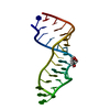 3dvzS S: Starting model for refinement |
|---|---|
| Similar structure data |
-Links
 PDBj
PDBj


- Assembly
| Deposited unit | 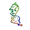
| ||||||||||||
|---|---|---|---|---|---|---|---|---|---|---|---|---|---|
| 1 |
| ||||||||||||
| Unit cell |
|
-Components
| #1: RNA chain | Mass: 8744.255 Da / Num. of mol.: 1 Source method: isolated from a genetically manipulated source Source: (gene. exp.) Homo sapiens (human) / Production host:  |
|---|---|
| #2: Chemical | ChemComp-SO4 /   Mass: 96.063 Da / Num. of mol.: 1 / Source method: obtained synthetically / Formula: SO4 Mass: 96.063 Da / Num. of mol.: 1 / Source method: obtained synthetically / Formula: SO4 |
| #3: Chemical | ChemComp-DOD /   Mass: 18.015 Da / Num. of mol.: 80 / Source method: isolated from a natural source / Formula: D2O / Feature type: SUBJECT OF INVESTIGATION Mass: 18.015 Da / Num. of mol.: 80 / Source method: isolated from a natural source / Formula: D2O / Feature type: SUBJECT OF INVESTIGATION |
| Has ligand of interest | Y |
-Experimental details
-Experiment
| Experiment |
|
|---|
- Sample preparation
| Crystal | Density Matthews: 1.9 Å3/Da / Density % sol: 35.37 % |
|---|---|
| Crystal grow | Temperature: 291.15 K / Method: vapor diffusion, sitting drop / pH: 7 Details: 0.233 mM RNA, 1.0 M ammonium sulfate, 2 mM magnesium chloride, 2 mM manganese chloride, 16.5 mM -3-(N-morpholino)propanesulfonic acid. |
-Data collection
| Diffraction |
| ||||||||||||||||||||||||
|---|---|---|---|---|---|---|---|---|---|---|---|---|---|---|---|---|---|---|---|---|---|---|---|---|---|
| Diffraction source |
| ||||||||||||||||||||||||
| Detector |
| ||||||||||||||||||||||||
| Radiation |
| ||||||||||||||||||||||||
| Radiation wavelength |
| ||||||||||||||||||||||||
| Reflection | Biso Wilson estimate: 8.3 Å2 / Entry-ID: 7UCR
| ||||||||||||||||||||||||
| Reflection shell |
|

- Processing
| Software |
| ||||||||||||||||||||||||||||||||||||||||||||||||||||||||||||||||||||||||||||||||||||||||||||||||||||||||||||||||||||||||||||||||||||||||||||||||||||||||||||||||||||||||||||||||||||||
|---|---|---|---|---|---|---|---|---|---|---|---|---|---|---|---|---|---|---|---|---|---|---|---|---|---|---|---|---|---|---|---|---|---|---|---|---|---|---|---|---|---|---|---|---|---|---|---|---|---|---|---|---|---|---|---|---|---|---|---|---|---|---|---|---|---|---|---|---|---|---|---|---|---|---|---|---|---|---|---|---|---|---|---|---|---|---|---|---|---|---|---|---|---|---|---|---|---|---|---|---|---|---|---|---|---|---|---|---|---|---|---|---|---|---|---|---|---|---|---|---|---|---|---|---|---|---|---|---|---|---|---|---|---|---|---|---|---|---|---|---|---|---|---|---|---|---|---|---|---|---|---|---|---|---|---|---|---|---|---|---|---|---|---|---|---|---|---|---|---|---|---|---|---|---|---|---|---|---|---|---|---|---|---|
| Refinement | SU ML: 0.1008 / Cross valid method: FREE R-VALUE / Method to determine structure:
| ||||||||||||||||||||||||||||||||||||||||||||||||||||||||||||||||||||||||||||||||||||||||||||||||||||||||||||||||||||||||||||||||||||||||||||||||||||||||||||||||||||||||||||||||||||||
| Refinement step | Cycle: LAST / Resolution: 1→16.05 Å
| ||||||||||||||||||||||||||||||||||||||||||||||||||||||||||||||||||||||||||||||||||||||||||||||||||||||||||||||||||||||||||||||||||||||||||||||||||||||||||||||||||||||||||||||||||||||
| Refine LS restraints |
| ||||||||||||||||||||||||||||||||||||||||||||||||||||||||||||||||||||||||||||||||||||||||||||||||||||||||||||||||||||||||||||||||||||||||||||||||||||||||||||||||||||||||||||||||||||||
| LS refinement shell |
|
