 Movie
Movie Controller

[English] 日本語
 Yorodumi
- PDB-7ua6: Horse liver alcohol dehydrogenase with NAD and trifluoroethanol at 25K -
+ Open data

- Basic information
| Entry | Database: PDB / ID: 7ua6 | |||||||||
|---|---|---|---|---|---|---|---|---|---|---|
| Title | Horse liver alcohol dehydrogenase with NAD and trifluoroethanol at 25K | |||||||||
 Components Components | Alcohol dehydrogenase E chain | |||||||||
 Keywords Keywords | METAL BINDING PROTEIN / Oxidoreductase / alcohol dehydrogenase liver | |||||||||
| Function / homology |  Function and homology information Function and homology informationall-trans-retinol dehydrogenase (NAD+) activity / alcohol dehydrogenase / retinoic acid metabolic process / retinol metabolic process / zinc ion binding / cytosol Similarity search - Function | |||||||||
| Biological species |  | |||||||||
| Method |  X-RAY DIFFRACTION / SYNCHROTRON / MOLECULAR REPLACEMENT / Resolution: 1.1 Å X-RAY DIFFRACTION / SYNCHROTRON / MOLECULAR REPLACEMENT / Resolution: 1.1 Å | |||||||||
 Authors Authors | Plapp, B.V. / Gakhar, L. | |||||||||
| Funding support |  United States, 2items United States, 2items
| |||||||||
 Citation Citation | Journal: Acta Crystallogr.,Sect.D / Year: 2022 Title: Dependence of crystallographic atomic displacement factors on temperature (25-150 K) for complexes of horse liver alcohol dehydrogenases Authors: Plapp, B.V. / Gakhar, L. / Subramanian, R. #1: Journal: Biochemistry / Year: 2012Title: Atomic-resolution structures of horse liver alcohol dehydrogenase with NAD(+) and fluoroalcohols define strained Michaelis complexes. Authors: Plapp, B.V. / Ramaswamy, S. | |||||||||
| History |
|
- Structure visualization
| Structure viewer | Molecule: MolmilJmol/JSmol |
|---|
- Downloads & links
-Download
| PDBx/mmCIF format | 7ua6.cif.gz | 345.3 KB | Display | PDBx/mmCIF format |
|---|---|---|---|---|
| PDB format | pdb7ua6.ent.gz | 279.4 KB | Display | PDB format |
| PDBx/mmJSON format | 7ua6.json.gz | Tree view | PDBx/mmJSON format | |
| Others |  Other downloads Other downloads |
-Validation report
| Arichive directory | https://data.pdbj.org/pub/pdb/validation_reports/ua/7ua6ftp://data.pdbj.org/pub/pdb/validation_reports/ua/7ua6 | HTTPS FTP |
|---|
-Related structure data
| Related structure data | 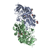 7uc9C  7ucaC  7ucuC 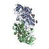 7uddC  7udeC  7udrC  7uecC  7ueeC  7uefC  7ueiC  7uejC  7uhvC  7uhwC  7uhxC  4dxhS S: Starting model for refinement C: citing same article ( |
|---|---|
| Similar structure data | |
| Experimental dataset #1 | Data reference: 10.15785/SBGRID/886 / Data set type: diffraction image data |
-Links
 PDBj
PDBj

- Assembly
| Deposited unit | 
| ||||||||
|---|---|---|---|---|---|---|---|---|---|
| 1 |
| ||||||||
| Unit cell |
|
-Components
-Protein , 1 types, 2 molecules AB
| #1: Protein | Mass: 39853.273 Da / Num. of mol.: 2 / Source method: isolated from a natural source / Source: (natural) |
|---|
-Non-polymers , 5 types, 887 molecules 
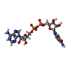
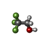


| #2: Chemical | ChemComp-ZN /  Mass: 65.409 Da / Num. of mol.: 4 / Source method: obtained synthetically / Formula: Zn / Feature type: SUBJECT OF INVESTIGATION Mass: 65.409 Da / Num. of mol.: 4 / Source method: obtained synthetically / Formula: Zn / Feature type: SUBJECT OF INVESTIGATION#3: Chemical |  Mass: 663.425 Da / Num. of mol.: 2 / Source method: obtained synthetically / Formula: C21H27N7O14P2 / Feature type: SUBJECT OF INVESTIGATION Mass: 663.425 Da / Num. of mol.: 2 / Source method: obtained synthetically / Formula: C21H27N7O14P2 / Feature type: SUBJECT OF INVESTIGATION#4: Chemical |  Mass: 100.040 Da / Num. of mol.: 2 / Source method: obtained synthetically / Formula: C2H3F3O / Feature type: SUBJECT OF INVESTIGATION Mass: 100.040 Da / Num. of mol.: 2 / Source method: obtained synthetically / Formula: C2H3F3O / Feature type: SUBJECT OF INVESTIGATION#5: Chemical | ChemComp-MRD / (  Mass: 118.174 Da / Num. of mol.: 4 / Source method: obtained synthetically / Formula: C6H14O2 / Comment: precipitant*YM Mass: 118.174 Da / Num. of mol.: 4 / Source method: obtained synthetically / Formula: C6H14O2 / Comment: precipitant*YM#6: Water | ChemComp-HOH / | Mass: 18.015 Da / Num. of mol.: 875 / Source method: isolated from a natural source / Formula: H2O |
|---|
-Details
| Has ligand of interest | Y |
|---|
-Experimental details
-Experiment
| Experiment | Method: X-RAY DIFFRACTION / Number of used crystals: 1 |
|---|
- Sample preparation
| Crystal | Density Matthews: 2.25 Å3/Da / Density % sol: 45 % / Description: block |
|---|---|
| Crystal grow | Temperature: 278 K / Method: microdialysis / pH: 7 Details: 50 MM AMMONIUM N-[TRIS(HYDROXYMETHYL) METHYL]-2-AMINOETHANE SULFONATE, PH 6.7 (AT 25 C), 0.25 MM EDTA, 10 MG/ML PROTEIN, 1 MM NAD+, 100 MM 2,2,2-TRIFLUOROETHANOL, 12 TO 25 % 2-METHYL-2,4-PENTANEDIOL, |
-Data collection
| Diffraction | Mean temperature: 25 K / Ambient temp details: He cryostat / Serial crystal experiment: N | ||||||||||||||||||||||||||||||||||||||||||||||||||||||||||||||||||||||||||||||||||||||||
|---|---|---|---|---|---|---|---|---|---|---|---|---|---|---|---|---|---|---|---|---|---|---|---|---|---|---|---|---|---|---|---|---|---|---|---|---|---|---|---|---|---|---|---|---|---|---|---|---|---|---|---|---|---|---|---|---|---|---|---|---|---|---|---|---|---|---|---|---|---|---|---|---|---|---|---|---|---|---|---|---|---|---|---|---|---|---|---|---|---|
| Diffraction source | Source: SYNCHROTRON / Site: APS / Beamline: 19-ID / Wavelength: 0.9184 Å | ||||||||||||||||||||||||||||||||||||||||||||||||||||||||||||||||||||||||||||||||||||||||
| Detector | Type: ADSC QUANTUM 315r / Detector: CCD / Date: Jun 24, 2009 Details: ROSENBAUM ROCK VERTICAL FOCUSINGMIRROR WITH PT, GLASS, PD LANES | ||||||||||||||||||||||||||||||||||||||||||||||||||||||||||||||||||||||||||||||||||||||||
| Radiation | Monochromator: ROSENBAUM ROCK HIGH RESOLUTION DOUBLE CRYSTAL MONOCHROMATOR, LN2 COOLED, SAGITTAL FOCUSSING 2ND MIRROR, SI(111) Protocol: SINGLE WAVELENGTH / Monochromatic (M) / Laue (L): M / Scattering type: x-ray | ||||||||||||||||||||||||||||||||||||||||||||||||||||||||||||||||||||||||||||||||||||||||
| Radiation wavelength | Wavelength: 0.9184 Å / Relative weight: 1 | ||||||||||||||||||||||||||||||||||||||||||||||||||||||||||||||||||||||||||||||||||||||||
| Reflection | Resolution: 1.1→19.93 Å / Num. obs: 281705 / % possible obs: 94.3 % / Redundancy: 4.06 % / Biso Wilson estimate: 9 Å2 / Rmerge(I) obs: 0.062 / Rpim(I) all: 0.027 / Rrim(I) all: 0.072 / Χ2: 1.19 / Net I/σ(I): 9.1 | ||||||||||||||||||||||||||||||||||||||||||||||||||||||||||||||||||||||||||||||||||||||||
| Reflection shell | Diffraction-ID: 1
|
- Processing
| Software |
| |||||||||||||||||||||||||||||||||||||||||||||||||||||||||||||||||
|---|---|---|---|---|---|---|---|---|---|---|---|---|---|---|---|---|---|---|---|---|---|---|---|---|---|---|---|---|---|---|---|---|---|---|---|---|---|---|---|---|---|---|---|---|---|---|---|---|---|---|---|---|---|---|---|---|---|---|---|---|---|---|---|---|---|---|
| Refinement | Method to determine structure: MOLECULAR REPLACEMENT Starting model: 4DXH Resolution: 1.1→19.93 Å / Cor.coef. Fo:Fc: 0.983 / Cor.coef. Fo:Fc free: 0.978 / SU B: 0.821 / SU ML: 0.017 / Cross valid method: THROUGHOUT / σ(F): 0 / ESU R: 0.025 / ESU R Free: 0.025 / Stereochemistry target values: MAXIMUM LIKELIHOOD Details: HYDROGENS HAVE BEEN ADDED IN THE RIDING POSITIONS U VALUES : REFINED INDIVIDUALLY
| |||||||||||||||||||||||||||||||||||||||||||||||||||||||||||||||||
| Solvent computation | Ion probe radii: 0.8 Å / Shrinkage radii: 0.8 Å / VDW probe radii: 1.2 Å / Solvent model: MASK | |||||||||||||||||||||||||||||||||||||||||||||||||||||||||||||||||
| Displacement parameters | Biso max: 61.1 Å2 / Biso mean: 13.843 Å2 / Biso min: 6.43 Å2
| |||||||||||||||||||||||||||||||||||||||||||||||||||||||||||||||||
| Refinement step | Cycle: final / Resolution: 1.1→19.93 Å
| |||||||||||||||||||||||||||||||||||||||||||||||||||||||||||||||||
| Refine LS restraints |
| |||||||||||||||||||||||||||||||||||||||||||||||||||||||||||||||||
| LS refinement shell | Resolution: 1.1→1.129 Å / Rfactor Rfree error: 0 / Total num. of bins used: 20
|
