 Movie
Movie Controller

[English] 日本語
 Yorodumi
- PDB-7u55: Crystal structure of Thermoplasmatales archaeon heliorhodopsin at... -
+ Open data

- Basic information
| Entry | Database: PDB / ID: 7u55 | ||||||
|---|---|---|---|---|---|---|---|
| Title | Crystal structure of Thermoplasmatales archaeon heliorhodopsin at pH 4.5 | ||||||
 Components Components | Heliorhodopsin | ||||||
 Keywords Keywords | MEMBRANE PROTEIN / microbial rhodopsin / retinal protein / heliorhodopsin / seven transmembrane protein | ||||||
| Function / homology | Heliorhodopsin / Heliorhodopsin / identical protein binding / membrane / DODECANE / RETINAL / Heliorhodopsin HeR Function and homology information Function and homology information | ||||||
| Biological species |  Thermoplasmatales archaeon SG8-52-1 (archaea) Thermoplasmatales archaeon SG8-52-1 (archaea) | ||||||
| Method |  X-RAY DIFFRACTION / SYNCHROTRON / MOLECULAR REPLACEMENT / Resolution: 1.97 Å X-RAY DIFFRACTION / SYNCHROTRON / MOLECULAR REPLACEMENT / Resolution: 1.97 Å | ||||||
 Authors Authors | Besaw, J.E. / De Guzman, P. / Miller, R.J.D. / Ernst, O.P. | ||||||
| Funding support |  Canada, 1items Canada, 1items
| ||||||
 Citation Citation | Journal: Sci Rep / Year: 2022 Title: Low pH structure of heliorhodopsin reveals chloride binding site and intramolecular signaling pathway. Authors: Besaw, J.E. / Reichenwallner, J. / De Guzman, P. / Tucs, A. / Kuo, A. / Morizumi, T. / Tsuda, K. / Sljoka, A. / Miller, R.J.D. / Ernst, O.P. | ||||||
| History |
|
- Structure visualization
| Structure viewer | Molecule: MolmilJmol/JSmol |
|---|
- Downloads & links
-Download
| PDBx/mmCIF format | 7u55.cif.gz | 72.7 KB | Display | PDBx/mmCIF format |
|---|---|---|---|---|
| PDB format | pdb7u55.ent.gz | 47.3 KB | Display | PDB format |
| PDBx/mmJSON format | 7u55.json.gz | Tree view | PDBx/mmJSON format | |
| Others |  Other downloads Other downloads |
-Validation report
| Summary document | 7u55_validation.pdf.gz | 649.8 KB | Display | wwPDB validaton report |
|---|---|---|---|---|
| Full document | 7u55_full_validation.pdf.gz | 652.9 KB | Display | |
| Data in XML | 7u55_validation.xml.gz | 11.7 KB | Display | |
| Data in CIF | 7u55_validation.cif.gz | 15.2 KB | Display | |
| Arichive directory | https://data.pdbj.org/pub/pdb/validation_reports/u5/7u55ftp://data.pdbj.org/pub/pdb/validation_reports/u5/7u55 | HTTPS FTP |
-Related structure data
| Related structure data | 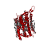 6is6S S: Starting model for refinement |
|---|---|
| Similar structure data |
-Links
 PDBj
PDBj


- Assembly
| Deposited unit | 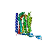
| ||||||||||||
|---|---|---|---|---|---|---|---|---|---|---|---|---|---|
| 1 | 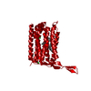
| ||||||||||||
| Unit cell |
|
-Components
| #1: Protein | Mass: 29886.301 Da / Num. of mol.: 1 Source method: isolated from a genetically manipulated source Source: (gene. exp.) Thermoplasmatales archaeon SG8-52-1 (archaea)Gene: AYK20_03510 / Production host:  | ||||||||
|---|---|---|---|---|---|---|---|---|---|
| #2: Chemical | ChemComp-RET /   Mass: 284.436 Da / Num. of mol.: 1 / Source method: obtained synthetically / Formula: C20H28O Mass: 284.436 Da / Num. of mol.: 1 / Source method: obtained synthetically / Formula: C20H28O | ||||||||
| #3: Chemical |   Mass: 35.453 Da / Num. of mol.: 2 / Source method: obtained synthetically / Formula: Cl Mass: 35.453 Da / Num. of mol.: 2 / Source method: obtained synthetically / Formula: Cl#4: Chemical | ChemComp-D12 / |   Mass: 170.335 Da / Num. of mol.: 1 / Source method: obtained synthetically / Formula: C12H26 Mass: 170.335 Da / Num. of mol.: 1 / Source method: obtained synthetically / Formula: C12H26#5: Water | ChemComp-HOH / |  Mass: 18.015 Da / Num. of mol.: 37 / Source method: isolated from a natural source / Formula: H2O Mass: 18.015 Da / Num. of mol.: 37 / Source method: isolated from a natural source / Formula: H2OHas ligand of interest | N | Has protein modification | Y | |
-Experimental details
-Experiment
| Experiment | Method: X-RAY DIFFRACTION / Number of used crystals: 1 |
|---|
- Sample preparation
| Crystal | Density Matthews: 2.04 Å3/Da / Density % sol: 39.82 % / Description: Diamond |
|---|---|
| Crystal grow | Temperature: 307.15 K / Method: vapor diffusion, hanging drop / pH: 4.5 Details: Bicelle crystallization with 5 mg/mL protein in 8% bicelle (2.8:1 DMPC:CHAPSO). Crystallization condition is 26% polyethylene glycol 3350, 0.1 M sodium phosphate monobasic monohydrate at pH ...Details: Bicelle crystallization with 5 mg/mL protein in 8% bicelle (2.8:1 DMPC:CHAPSO). Crystallization condition is 26% polyethylene glycol 3350, 0.1 M sodium phosphate monobasic monohydrate at pH 4.5, 0.28 M ammonium sulfate, 0.18 M 1,6-hexanediol |
-Data collection
| Diffraction | Mean temperature: 100 K / Serial crystal experiment: N |
|---|---|
| Diffraction source | Source: SYNCHROTRON / Site: APS  / Beamline: 23-ID-B / Wavelength: 1.033 Å / Beamline: 23-ID-B / Wavelength: 1.033 Å |
| Detector | Type: DECTRIS EIGER X 16M / Detector: PIXEL / Date: Nov 9, 2019 |
| Radiation | Protocol: SINGLE WAVELENGTH / Monochromatic (M) / Laue (L): M / Scattering type: x-ray |
| Radiation wavelength | Wavelength: 1.033 Å / Relative weight: 1 |
| Reflection | Resolution: 1.97→47.94 Å / Num. obs: 16754 / % possible obs: 94.4 % / Redundancy: 3.3 % / Biso Wilson estimate: 22.85 Å2 / CC1/2: 0.98 / Net I/σ(I): 4.3 |
| Reflection shell | Resolution: 1.97→2.02 Å / Redundancy: 2.2 % / Mean I/σ(I) obs: 1.3 / Num. unique obs: 1100 / CC1/2: 0.72 / % possible all: 89.2 |
- Processing
| Software |
| |||||||||||||||||||||||||||||||||||||||||||||||||||||||||||||||||||||||||||||||||||||||||||
|---|---|---|---|---|---|---|---|---|---|---|---|---|---|---|---|---|---|---|---|---|---|---|---|---|---|---|---|---|---|---|---|---|---|---|---|---|---|---|---|---|---|---|---|---|---|---|---|---|---|---|---|---|---|---|---|---|---|---|---|---|---|---|---|---|---|---|---|---|---|---|---|---|---|---|---|---|---|---|---|---|---|---|---|---|---|---|---|---|---|---|---|---|
| Refinement | Method to determine structure: MOLECULAR REPLACEMENT Starting model: 6is6 Resolution: 1.97→47.94 Å / SU ML: 0.1674 / Cross valid method: FREE R-VALUE / σ(F): 1.34 / Phase error: 20.3999 Stereochemistry target values: GeoStd + Monomer Library + CDL v1.2
| |||||||||||||||||||||||||||||||||||||||||||||||||||||||||||||||||||||||||||||||||||||||||||
| Solvent computation | Shrinkage radii: 0.9 Å / VDW probe radii: 1.11 Å / Solvent model: FLAT BULK SOLVENT MODEL | |||||||||||||||||||||||||||||||||||||||||||||||||||||||||||||||||||||||||||||||||||||||||||
| Displacement parameters | Biso mean: 28.56 Å2 | |||||||||||||||||||||||||||||||||||||||||||||||||||||||||||||||||||||||||||||||||||||||||||
| Refinement step | Cycle: LAST / Resolution: 1.97→47.94 Å
| |||||||||||||||||||||||||||||||||||||||||||||||||||||||||||||||||||||||||||||||||||||||||||
| Refine LS restraints |
| |||||||||||||||||||||||||||||||||||||||||||||||||||||||||||||||||||||||||||||||||||||||||||
| LS refinement shell |
|
