 Movie
Movie Controller


+ Open data

- Basic information
| Entry | Database: PDB / ID: 7u4g | ||||||||||||
|---|---|---|---|---|---|---|---|---|---|---|---|---|---|
| Title | Neuraminidase from influenza virus A/Shandong/9/1993(H3N2) | ||||||||||||
 Components Components | Neuraminidase | ||||||||||||
 Keywords Keywords | VIRAL PROTEIN / Hydrolase / neuraminidase / influenza | ||||||||||||
| Function / homology |  Function and homology information Function and homology information: / : / : / exo-alpha-sialidase / viral budding from plasma membrane / carbohydrate metabolic process / host cell plasma membrane / virion membrane / membrane / metal ion binding Similarity search - Function | ||||||||||||
| Biological species |   Influenza A virus Influenza A virus | ||||||||||||
| Method |  X-RAY DIFFRACTION / SYNCHROTRON / MOLECULAR REPLACEMENT / Resolution: 1.65 Å X-RAY DIFFRACTION / SYNCHROTRON / MOLECULAR REPLACEMENT / Resolution: 1.65 Å | ||||||||||||
 Authors Authors | Lei, R. / Hernandez Garcia, A. | ||||||||||||
| Funding support |  United States, 3items United States, 3items
| ||||||||||||
 Citation Citation | Journal: Nat Commun / Year: 2022 Title: Prevalence and mechanisms of evolutionary contingency in human influenza H3N2 neuraminidase. Authors: Lei, R. / Tan, T.J.C. / Hernandez Garcia, A. / Wang, Y. / Diefenbacher, M. / Teo, C. / Gopan, G. / Tavakoli Dargani, Z. / Teo, Q.W. / Graham, C.S. / Brooke, C.B. / Nair, S.K. / Wu, N.C. | ||||||||||||
| History |
|
- Structure visualization
| Structure viewer | Molecule: MolmilJmol/JSmol |
|---|
- Downloads & links
-Download
| PDBx/mmCIF format | 7u4g.cif.gz | 104.7 KB | Display | PDBx/mmCIF format |
|---|---|---|---|---|
| PDB format | pdb7u4g.ent.gz | 75.5 KB | Display | PDB format |
| PDBx/mmJSON format | 7u4g.json.gz | Tree view | PDBx/mmJSON format | |
| Others |  Other downloads Other downloads |
-Validation report
| Arichive directory | https://data.pdbj.org/pub/pdb/validation_reports/u4/7u4gftp://data.pdbj.org/pub/pdb/validation_reports/u4/7u4g | HTTPS FTP |
|---|
-Related structure data
| Related structure data | 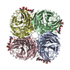 7u4eC 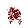 7u4fC 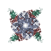 2aepS S: Starting model for refinement C: citing same article ( |
|---|---|
| Similar structure data |
-Links
 PDBj
PDBj

- Assembly
| Deposited unit | 
| ||||||||
|---|---|---|---|---|---|---|---|---|---|
| 1 | 
| ||||||||
| Unit cell |
| ||||||||
| Components on special symmetry positions |
|
-Components
-Protein , 1 types, 1 molecules A
| #1: Protein | Mass: 52186.840 Da / Num. of mol.: 1 Source method: isolated from a genetically manipulated source Source: (gene. exp.) Influenza A virus (A/Shandong/9/1993(H3N2))Gene: NA / Production host: unidentified baculovirus / References: UniProt: Q98815, exo-alpha-sialidase |
|---|
-Sugars , 4 types, 4 molecules 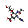
| #2: Polysaccharide | alpha-D-mannopyranose-(1-2)-alpha-D-mannopyranose-(1-3)-[alpha-D-mannopyranose-(1-6)]beta-D- ...alpha-D-mannopyranose-(1-2)-alpha-D-mannopyranose-(1-3)-[alpha-D-mannopyranose-(1-6)]beta-D-mannopyranose-(1-4)-2-acetamido-2-deoxy-beta-D-glucopyranose-(1-4)-2-acetamido-2-deoxy-beta-D-glucopyranose Source method: isolated from a genetically manipulated source |
|---|---|
| #3: Polysaccharide | alpha-D-mannopyranose-(1-4)-2-acetamido-2-deoxy-beta-D-glucopyranose Source method: isolated from a genetically manipulated source |
| #4: Polysaccharide | 2-acetamido-2-deoxy-beta-D-glucopyranose-(1-4)-2-acetamido-2-deoxy-beta-D-glucopyranose Source method: isolated from a genetically manipulated source |
| #5: Sugar | ChemComp-ZMR /  Type: D-saccharide / Mass: 332.310 Da / Num. of mol.: 1 / Source method: obtained synthetically / Formula: C12H20N4O7 / Comment: medication, antivirus, inhibitor*YM Type: D-saccharide / Mass: 332.310 Da / Num. of mol.: 1 / Source method: obtained synthetically / Formula: C12H20N4O7 / Comment: medication, antivirus, inhibitor*YM |
-Non-polymers , 2 types, 318 molecules 

| #6: Chemical | ChemComp-CA /  Mass: 40.078 Da / Num. of mol.: 1 / Source method: obtained synthetically / Formula: Ca Mass: 40.078 Da / Num. of mol.: 1 / Source method: obtained synthetically / Formula: Ca |
|---|---|
| #7: Water | ChemComp-HOH / Mass: 18.015 Da / Num. of mol.: 317 / Source method: isolated from a natural source / Formula: H2O |
-Details
| Has ligand of interest | N |
|---|---|
| Has protein modification | Y |
-Experimental details
-Experiment
| Experiment | Method: X-RAY DIFFRACTION / Number of used crystals: 1 |
|---|
- Sample preparation
| Crystal | Density Matthews: 2.19 Å3/Da / Density % sol: 43.95 % |
|---|---|
| Crystal grow | Temperature: 291.15 K / Method: vapor diffusion, sitting drop / Details: 8% PEG-8000, 0.1 M Tris pH 8.5 |
-Data collection
| Diffraction | Mean temperature: 100 K / Serial crystal experiment: N |
|---|---|
| Diffraction source | Source: SYNCHROTRON / Site: APS / Beamline: 21-ID-D / Wavelength: 1.1272 Å |
| Detector | Type: DECTRIS EIGER X 9M / Detector: PIXEL / Date: Nov 26, 2021 |
| Radiation | Protocol: SINGLE WAVELENGTH / Monochromatic (M) / Laue (L): M / Scattering type: x-ray |
| Radiation wavelength | Wavelength: 1.1272 Å / Relative weight: 1 |
| Reflection | Resolution: 1.645→76.34 Å / Num. obs: 54981 / % possible obs: 96.8 % / Redundancy: 9.5 % / CC1/2: 0.998 / Rmerge(I) obs: 0.127 / Rrim(I) all: 0.138 / Net I/σ(I): 11.5 |
| Reflection shell | Resolution: 1.645→1.65 Å / Rmerge(I) obs: 0.774 / Mean I/σ(I) obs: 2.1 / CC1/2: 0.895 / Rrim(I) all: 0.834 |
- Processing
| Software |
| |||||||||||||||||||||||||||||||||||||||||||||
|---|---|---|---|---|---|---|---|---|---|---|---|---|---|---|---|---|---|---|---|---|---|---|---|---|---|---|---|---|---|---|---|---|---|---|---|---|---|---|---|---|---|---|---|---|---|---|
| Refinement | Method to determine structure: MOLECULAR REPLACEMENT Starting model: 2AEP Resolution: 1.65→25 Å / Cor.coef. Fo:Fc: 0.962 / Cor.coef. Fo:Fc free: 0.946 / SU B: 3.338 / SU ML: 0.098 / Cross valid method: THROUGHOUT / σ(F): 0 / ESU R: 0.097 / ESU R Free: 0.094 / Stereochemistry target values: MAXIMUM LIKELIHOOD Details: HYDROGENS HAVE BEEN USED IF PRESENT IN THE INPUT U VALUES : REFINED INDIVIDUALLY
| |||||||||||||||||||||||||||||||||||||||||||||
| Solvent computation | Ion probe radii: 0.8 Å / Shrinkage radii: 0.8 Å / VDW probe radii: 1.2 Å / Solvent model: MASK | |||||||||||||||||||||||||||||||||||||||||||||
| Displacement parameters | Biso max: 64.35 Å2 / Biso mean: 19.373 Å2 / Biso min: 7.94 Å2
| |||||||||||||||||||||||||||||||||||||||||||||
| Refinement step | Cycle: final / Resolution: 1.65→25 Å
| |||||||||||||||||||||||||||||||||||||||||||||
| Refine LS restraints |
| |||||||||||||||||||||||||||||||||||||||||||||
| LS refinement shell | Resolution: 1.65→1.693 Å / Rfactor Rfree error: 0 / Total num. of bins used: 20
|
