 Movie
Movie Controller


+ Open data

- Basic information
| Entry | Database: PDB / ID: 7u3h | ||||||
|---|---|---|---|---|---|---|---|
| Title | GID4 in complex with compound 7 | ||||||
 Components Components | Glucose-induced degradation protein 4 homolog | ||||||
 Keywords Keywords | PEPTIDE BINDING PROTEIN/INHIBITOR / Complex / Inhibitor / Protein degradation / PEPTIDE BINDING PROTEIN / PEPTIDE BINDING PROTEIN-INHIBITOR complex | ||||||
| Function / homology | Vacuolar import/degradation protein Vid24 / Vacuolar import and degradation protein / ubiquitin ligase complex / Regulation of pyruvate metabolism / ubiquitin protein ligase activity / proteasome-mediated ubiquitin-dependent protein catabolic process / cytosol / Chem-L6O / Glucose-induced degradation protein 4 homolog Function and homology information Function and homology information | ||||||
| Biological species |  Homo sapiens (human) Homo sapiens (human) | ||||||
| Method |  X-RAY DIFFRACTION / SYNCHROTRON / MOLECULAR REPLACEMENT / molecular replacement / Resolution: 1.798 Å X-RAY DIFFRACTION / SYNCHROTRON / MOLECULAR REPLACEMENT / molecular replacement / Resolution: 1.798 Å | ||||||
 Authors Authors | Chana, C.K. / Sicheri, F. | ||||||
| Funding support |  Canada, 1items Canada, 1items
| ||||||
 Citation Citation | Journal: J.Med.Chem. / Year: 2022 Title: Discovery and Structural Characterization of Small Molecule Binders of the Human CTLH E3 Ligase Subunit GID4. Authors: Chana, C.K. / Maisonneuve, P. / Posternak, G. / Grinberg, N.G.A. / Poirson, J. / Ona, S.M. / Ceccarelli, D.F. / Mader, P. / St-Cyr, D.J. / Pau, V. / Kurinov, I. / Tang, X. / Deng, D. / Cui, ...Authors: Chana, C.K. / Maisonneuve, P. / Posternak, G. / Grinberg, N.G.A. / Poirson, J. / Ona, S.M. / Ceccarelli, D.F. / Mader, P. / St-Cyr, D.J. / Pau, V. / Kurinov, I. / Tang, X. / Deng, D. / Cui, W. / Su, W. / Kuai, L. / Soll, R. / Tyers, M. / Rost, H.L. / Batey, R.A. / Taipale, M. / Gingras, A.C. / Sicheri, F. | ||||||
| History |
|
- Structure visualization
| Structure viewer | Molecule: MolmilJmol/JSmol |
|---|
- Downloads & links
-Download
| PDBx/mmCIF format | 7u3h.cif.gz | 86.8 KB | Display | PDBx/mmCIF format |
|---|---|---|---|---|
| PDB format | pdb7u3h.ent.gz | 62.2 KB | Display | PDB format |
| PDBx/mmJSON format | 7u3h.json.gz | Tree view | PDBx/mmJSON format | |
| Others |  Other downloads Other downloads |
-Validation report
| Arichive directory | https://data.pdbj.org/pub/pdb/validation_reports/u3/7u3hftp://data.pdbj.org/pub/pdb/validation_reports/u3/7u3h | HTTPS FTP |
|---|
-Related structure data
| Related structure data | 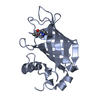 7u3eC 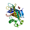 7u3fC 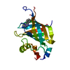 7u3gC 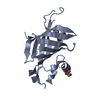 7u3iC 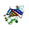 7u3jC 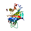 7u3kC 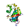 7u3lC 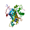 6ccrS S: Starting model for refinement C: citing same article ( |
|---|---|
| Similar structure data |
-Links
 PDBj
PDBj- Assembly
| Deposited unit | 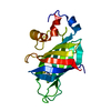
| ||||||||
|---|---|---|---|---|---|---|---|---|---|
| 1 |
| ||||||||
| Unit cell |
|
-Components
| #1: Protein | Mass: 19604.777 Da / Num. of mol.: 1 Source method: isolated from a genetically manipulated source Source: (gene. exp.) Homo sapiens (human) / Gene: GID4, C17orf39, VID24 / Plasmid: PET28-MHL / Production host:  |
|---|---|
| #2: Chemical | ChemComp-L6O / (  Mass: 210.271 Da / Num. of mol.: 1 / Source method: obtained synthetically / Formula: C10H11FN2S / Feature type: SUBJECT OF INVESTIGATION Mass: 210.271 Da / Num. of mol.: 1 / Source method: obtained synthetically / Formula: C10H11FN2S / Feature type: SUBJECT OF INVESTIGATION |
| #3: Water | ChemComp-HOH /  Mass: 18.015 Da / Num. of mol.: 86 / Source method: isolated from a natural source / Formula: H2O Mass: 18.015 Da / Num. of mol.: 86 / Source method: isolated from a natural source / Formula: H2O |
| Has ligand of interest | Y |
-Experimental details
-Experiment
| Experiment | Method: X-RAY DIFFRACTION / Number of used crystals: 1 |
|---|
- Sample preparation
| Crystal | Density Matthews: 1.95 Å3/Da / Density % sol: 36.98 % |
|---|---|
| Crystal grow | Temperature: 293 K / Method: vapor diffusion, hanging drop / pH: 6.5 Details: 14% PEG3350, 0.1 M Bis-tris, 0.2 M ammonium formate |
-Data collection
| Diffraction | Mean temperature: 100 K / Serial crystal experiment: N | ||||||||||||||||||||||||||||||||||||||||||||||||||||||||||||||||||||||||||||||||||||||||||||||||||||
|---|---|---|---|---|---|---|---|---|---|---|---|---|---|---|---|---|---|---|---|---|---|---|---|---|---|---|---|---|---|---|---|---|---|---|---|---|---|---|---|---|---|---|---|---|---|---|---|---|---|---|---|---|---|---|---|---|---|---|---|---|---|---|---|---|---|---|---|---|---|---|---|---|---|---|---|---|---|---|---|---|---|---|---|---|---|---|---|---|---|---|---|---|---|---|---|---|---|---|---|---|---|
| Diffraction source | Source: SYNCHROTRON / Site: APS  / Beamline: 24-ID-C / Wavelength: 0.97918 Å / Beamline: 24-ID-C / Wavelength: 0.97918 Å | ||||||||||||||||||||||||||||||||||||||||||||||||||||||||||||||||||||||||||||||||||||||||||||||||||||
| Detector | Type: DECTRIS EIGER2 X 16M / Detector: PIXEL / Date: Jul 24, 2020 | ||||||||||||||||||||||||||||||||||||||||||||||||||||||||||||||||||||||||||||||||||||||||||||||||||||
| Radiation | Protocol: SINGLE WAVELENGTH / Monochromatic (M) / Laue (L): M / Scattering type: x-ray | ||||||||||||||||||||||||||||||||||||||||||||||||||||||||||||||||||||||||||||||||||||||||||||||||||||
| Radiation wavelength | Wavelength: 0.97918 Å / Relative weight: 1 | ||||||||||||||||||||||||||||||||||||||||||||||||||||||||||||||||||||||||||||||||||||||||||||||||||||
| Reflection | Resolution: 1.798→49.08 Å / Num. obs: 13855 / % possible obs: 97.1 % / Redundancy: 3.486 % / Biso Wilson estimate: 35.47 Å2 / CC1/2: 0.999 / Rmerge(I) obs: 0.052 / Rrim(I) all: 0.062 / Χ2: 0.799 / Net I/σ(I): 13.56 | ||||||||||||||||||||||||||||||||||||||||||||||||||||||||||||||||||||||||||||||||||||||||||||||||||||
| Reflection shell | Diffraction-ID: 1
|
-Phasing
| Phasing | Method: molecular replacement | |||||||||
|---|---|---|---|---|---|---|---|---|---|---|
| Phasing MR |
|
- Processing
| Software |
| ||||||||||||||||||||||||||||||||||||||||||||||||||||||||||||||||||||||||||||||||||||||||||||||||||||
|---|---|---|---|---|---|---|---|---|---|---|---|---|---|---|---|---|---|---|---|---|---|---|---|---|---|---|---|---|---|---|---|---|---|---|---|---|---|---|---|---|---|---|---|---|---|---|---|---|---|---|---|---|---|---|---|---|---|---|---|---|---|---|---|---|---|---|---|---|---|---|---|---|---|---|---|---|---|---|---|---|---|---|---|---|---|---|---|---|---|---|---|---|---|---|---|---|---|---|---|---|---|
| Refinement | Method to determine structure: MOLECULAR REPLACEMENT Starting model: 6CCR Resolution: 1.798→49.08 Å / SU ML: 0.25 / Cross valid method: THROUGHOUT / σ(F): 1.35 / Phase error: 27.34 / Stereochemistry target values: ML
| ||||||||||||||||||||||||||||||||||||||||||||||||||||||||||||||||||||||||||||||||||||||||||||||||||||
| Solvent computation | Shrinkage radii: 0.9 Å / VDW probe radii: 1.11 Å / Solvent model: FLAT BULK SOLVENT MODEL | ||||||||||||||||||||||||||||||||||||||||||||||||||||||||||||||||||||||||||||||||||||||||||||||||||||
| Displacement parameters | Biso max: 94.8 Å2 / Biso mean: 40.541 Å2 / Biso min: 16.32 Å2 | ||||||||||||||||||||||||||||||||||||||||||||||||||||||||||||||||||||||||||||||||||||||||||||||||||||
| Refinement step | Cycle: final / Resolution: 1.798→49.08 Å
| ||||||||||||||||||||||||||||||||||||||||||||||||||||||||||||||||||||||||||||||||||||||||||||||||||||
| LS refinement shell | Refine-ID: X-RAY DIFFRACTION / Rfactor Rfree error: 0
| ||||||||||||||||||||||||||||||||||||||||||||||||||||||||||||||||||||||||||||||||||||||||||||||||||||
| Refinement TLS params. | Method: refined / Refine-ID: X-RAY DIFFRACTION
| ||||||||||||||||||||||||||||||||||||||||||||||||||||||||||||||||||||||||||||||||||||||||||||||||||||
| Refinement TLS group |
|
