 Movie
Movie Controller


+ Open data

- Basic information
| Entry | Database: PDB / ID: 7u32 | |||||||||||||||||||||||||||||||||||||||||||||||||||||||||
|---|---|---|---|---|---|---|---|---|---|---|---|---|---|---|---|---|---|---|---|---|---|---|---|---|---|---|---|---|---|---|---|---|---|---|---|---|---|---|---|---|---|---|---|---|---|---|---|---|---|---|---|---|---|---|---|---|---|---|
| Title | MVV cleaved synaptic complex (CSC) intasome at 3.4 A resolution | |||||||||||||||||||||||||||||||||||||||||||||||||||||||||
 Components Components |
| |||||||||||||||||||||||||||||||||||||||||||||||||||||||||
 Keywords Keywords | VIRAL PROTEIN/DNA / Integrase-DNA complex / hydrolase / VIRAL PROTEIN / VIRAL PROTEIN-DNA complex | |||||||||||||||||||||||||||||||||||||||||||||||||||||||||
| Function / homology |  Function and homology information Function and homology informationdUTP diphosphatase / dUTP diphosphatase activity / nucleotide metabolic process / Hydrolases; Acting on peptide bonds (peptidases); Aspartic endopeptidases / ribonuclease H / exoribonuclease H / exoribonuclease H activity / DNA integration / viral genome integration into host DNA / establishment of integrated proviral latency ...dUTP diphosphatase / dUTP diphosphatase activity / nucleotide metabolic process / Hydrolases; Acting on peptide bonds (peptidases); Aspartic endopeptidases / ribonuclease H / exoribonuclease H / exoribonuclease H activity / DNA integration / viral genome integration into host DNA / establishment of integrated proviral latency / RNA-directed DNA polymerase / RNA stem-loop binding / RNA-directed DNA polymerase activity / RNA-DNA hybrid ribonuclease activity / Transferases; Transferring phosphorus-containing groups; Nucleotidyltransferases / viral capsid / DNA recombination / DNA-directed DNA polymerase / aspartic-type endopeptidase activity / Hydrolases; Acting on ester bonds / DNA-directed DNA polymerase activity / viral translational frameshifting / symbiont entry into host cell / proteolysis / DNA binding / zinc ion binding Similarity search - Function | |||||||||||||||||||||||||||||||||||||||||||||||||||||||||
| Biological species |  Visna/maedi virus EV1 KV1772 Visna/maedi virus EV1 KV1772synthetic construct (others) | |||||||||||||||||||||||||||||||||||||||||||||||||||||||||
| Method | ELECTRON MICROSCOPY / single particle reconstruction / cryo EM / Resolution: 3.46 Å | |||||||||||||||||||||||||||||||||||||||||||||||||||||||||
 Authors Authors | Shan, Z. / Pye, V.E. / Cherepanov, P. / Lyumkis, D. | |||||||||||||||||||||||||||||||||||||||||||||||||||||||||
| Funding support |  United States, United States,  United Kingdom, 5items United Kingdom, 5items
| |||||||||||||||||||||||||||||||||||||||||||||||||||||||||
 Citation Citation | Journal: Nat Commun / Year: 2022 Title: Multivalent interactions essential for lentiviral integrase function. Authors: Allison Ballandras-Colas / Vidya Chivukula / Dominika T Gruszka / Zelin Shan / Parmit K Singh / Valerie E Pye / Rebecca K McLean / Gregory J Bedwell / Wen Li / Andrea Nans / Nicola J Cook / ...Authors: Allison Ballandras-Colas / Vidya Chivukula / Dominika T Gruszka / Zelin Shan / Parmit K Singh / Valerie E Pye / Rebecca K McLean / Gregory J Bedwell / Wen Li / Andrea Nans / Nicola J Cook / Hind J Fadel / Eric M Poeschla / David J Griffiths / Javier Vargas / Ian A Taylor / Dmitry Lyumkis / Hasan Yardimci / Alan N Engelman / Peter Cherepanov /   Abstract: A multimer of retroviral integrase (IN) synapses viral DNA ends within a stable intasome nucleoprotein complex for integration into a host cell genome. Reconstitution of the intasome from the maedi- ...A multimer of retroviral integrase (IN) synapses viral DNA ends within a stable intasome nucleoprotein complex for integration into a host cell genome. Reconstitution of the intasome from the maedi-visna virus (MVV), an ovine lentivirus, revealed a large assembly containing sixteen IN subunits. Herein, we report cryo-EM structures of the lentiviral intasome prior to engagement of target DNA and following strand transfer, refined at 3.4 and 3.5 Å resolution, respectively. The structures elucidate details of the protein-protein and protein-DNA interfaces involved in lentiviral intasome formation. We show that the homomeric interfaces involved in IN hexadecamer formation and the α-helical configuration of the linker connecting the C-terminal and catalytic core domains are critical for MVV IN strand transfer activity in vitro and for virus infectivity. Single-molecule microscopy in conjunction with photobleaching reveals that the MVV intasome can bind a variable number, up to sixteen molecules, of the lentivirus-specific host factor LEDGF/p75. Concordantly, ablation of endogenous LEDGF/p75 results in gross redistribution of MVV integration sites in human and ovine cells. Our data confirm the importance of the expanded architecture observed in cryo-EM studies of lentiviral intasomes and suggest that this organization underlies multivalent interactions with chromatin for integration targeting to active genes. | |||||||||||||||||||||||||||||||||||||||||||||||||||||||||
| History |
|
- Structure visualization
| Structure viewer | Molecule: MolmilJmol/JSmol |
|---|
- Downloads & links
-Download
| PDBx/mmCIF format | 7u32.cif.gz | 793.3 KB | Display | PDBx/mmCIF format |
|---|---|---|---|---|
| PDB format | pdb7u32.ent.gz | 652.5 KB | Display | PDB format |
| PDBx/mmJSON format | 7u32.json.gz | Tree view | PDBx/mmJSON format | |
| Others |  Other downloads Other downloads |
-Validation report
| Arichive directory | https://data.pdbj.org/pub/pdb/validation_reports/u3/7u32ftp://data.pdbj.org/pub/pdb/validation_reports/u3/7u32 | HTTPS FTP |
|---|
-Related structure data
| Related structure data |  26322MC M: map data used to model this data C: citing same article ( |
|---|---|
| Similar structure data |
-Links
 PDBj
PDBj


- Assembly
| Deposited unit | 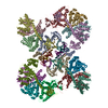
|
|---|---|
| 1 |
|
-Components
| #1: Protein | Mass: 32368.826 Da / Num. of mol.: 16 Source method: isolated from a genetically manipulated source Source: (gene. exp.) Visna/maedi virus EV1 KV1772 / Strain: KV1772 / Gene: pol / Production host:  References: UniProt: P35956, Transferases; Transferring phosphorus-containing groups; Nucleotidyltransferases, Hydrolases; Acting on ester bonds #2: DNA chain | Mass: 8943.719 Da / Num. of mol.: 2 / Source method: obtained synthetically / Source: (synth.) synthetic construct (others) #3: DNA chain | Mass: 8272.326 Da / Num. of mol.: 2 / Source method: obtained synthetically / Source: (synth.) synthetic construct (others) #4: Chemical | ChemComp-ZN /   Mass: 65.409 Da / Num. of mol.: 12 / Source method: obtained synthetically / Formula: Zn Mass: 65.409 Da / Num. of mol.: 12 / Source method: obtained synthetically / Formula: Zn#5: Chemical |   Mass: 40.078 Da / Num. of mol.: 2 / Source method: obtained synthetically / Formula: Ca Mass: 40.078 Da / Num. of mol.: 2 / Source method: obtained synthetically / Formula: CaHas ligand of interest | N | Has protein modification | N | |
|---|
-Experimental details
-Experiment
| Experiment | Method: ELECTRON MICROSCOPY |
|---|---|
| EM experiment | Aggregation state: PARTICLE / 3D reconstruction method: single particle reconstruction |
- Sample preparation
| Component | Name: MVV cleaved synaptic complex intasome / Type: COMPLEX / Entity ID: #1-#3 / Source: MULTIPLE SOURCES | |||||||||||||||||||||||||
|---|---|---|---|---|---|---|---|---|---|---|---|---|---|---|---|---|---|---|---|---|---|---|---|---|---|---|
| Molecular weight | Value: 0.48 kDa/nm / Experimental value: NO | |||||||||||||||||||||||||
| Source (natural) | Organism: Streptomyces griseus (bacteria) | |||||||||||||||||||||||||
| Source (recombinant) | Organism: | |||||||||||||||||||||||||
| Buffer solution | pH: 6.5 | |||||||||||||||||||||||||
| Buffer component |
| |||||||||||||||||||||||||
| Specimen | Embedding applied: NO / Shadowing applied: NO / Staining applied: NO / Vitrification applied: YES Details: MVV CSC intasomes, assembled and purified as previously described, were applied onto R1.2/1.3 gold UltrAufoil grids, Au 300 mesh (Quantifoil). Cryo-EM grids were prepared by manually ...Details: MVV CSC intasomes, assembled and purified as previously described, were applied onto R1.2/1.3 gold UltrAufoil grids, Au 300 mesh (Quantifoil). Cryo-EM grids were prepared by manually freezing using a manual plunger in cold room at 4C and stored in liquid nitrogen for future data acquisition. | |||||||||||||||||||||||||
| Specimen support | Grid material: GOLD / Grid mesh size: 300 divisions/in. / Grid type: Quantifoil R1.2/1.3 | |||||||||||||||||||||||||
| Vitrification | Instrument: HOMEMADE PLUNGER / Cryogen name: ETHANE Details: Cryo-EM grids were prepared by freezing using a manual plunger in cold room at 4C |
- Electron microscopy imaging
| Experimental equipment |  Model: Talos Arctica / Image courtesy: FEI Company |
|---|---|
| Microscopy | Model: FEI TALOS ARCTICA Details: The stage was tilted to 40 degrees during data collection to account for the preferential orientation of the sample within the vitreous ice. |
| Electron gun | Electron source:  FIELD EMISSION GUN / Accelerating voltage: 200 kV / Illumination mode: FLOOD BEAM FIELD EMISSION GUN / Accelerating voltage: 200 kV / Illumination mode: FLOOD BEAM |
| Electron lens | Mode: BRIGHT FIELD / Nominal magnification: 45000 X / Calibrated magnification: 54347 X / Nominal defocus max: 3000 nm / Nominal defocus min: 1000 nm / Cs: 2.7 mm / C2 aperture diameter: 70 µm / Alignment procedure: COMA FREE |
| Specimen holder | Cryogen: NITROGEN / Specimen holder model: FEI TITAN KRIOS AUTOGRID HOLDER |
| Image recording | Average exposure time: 10 sec. / Electron dose: 43.6 e/Å2 / Detector mode: COUNTING / Film or detector model: GATAN K2 SUMMIT (4k x 4k) / Num. of grids imaged: 1 / Num. of real images: 2295 |
| Image scans | Width: 3838 / Height: 3710 / Movie frames/image: 100 / Used frames/image: 1-100 |
- Processing
| Software | Name: PHENIX / Version: dev_4213: / Classification: refinement | ||||||||||||||||||||||||||||||||||||||||
|---|---|---|---|---|---|---|---|---|---|---|---|---|---|---|---|---|---|---|---|---|---|---|---|---|---|---|---|---|---|---|---|---|---|---|---|---|---|---|---|---|---|
| EM software |
| ||||||||||||||||||||||||||||||||||||||||
| CTF correction | Type: PHASE FLIPPING AND AMPLITUDE CORRECTION | ||||||||||||||||||||||||||||||||||||||||
| Particle selection | Num. of particles selected: 926176 | ||||||||||||||||||||||||||||||||||||||||
| Symmetry | Point symmetry: C2 (2 fold cyclic) | ||||||||||||||||||||||||||||||||||||||||
| 3D reconstruction | Resolution: 3.46 Å / Resolution method: FSC 0.143 CUT-OFF / Num. of particles: 147860 / Algorithm: FOURIER SPACE / Symmetry type: POINT | ||||||||||||||||||||||||||||||||||||||||
| Atomic model building | B value: 262 / Protocol: FLEXIBLE FIT / Space: REAL / Target criteria: CC Details: The STC model refined in study, with the tDNA removed, was docked into the CSC cryoEM map using UCSF Chimera. It was observed that there were some slight differences in some domain ...Details: The STC model refined in study, with the tDNA removed, was docked into the CSC cryoEM map using UCSF Chimera. It was observed that there were some slight differences in some domain positions, to address this, individual domains that were not well fitted to the map were docked as individual domains to achieve a best-fit starting model. Two (C2 related) NTDs (aa 1-35) were removed from the model due to lack of supporting map. Adjustments were made to the model interactively using Coot and the coordinates were subjected to real-space refinement in Phenix dev-4213-000 employing C2 NCS constraints. | ||||||||||||||||||||||||||||||||||||||||
| Refine LS restraints |
|
