 Movie
Movie Controller


+ Open data

- Basic information
| Entry | Database: PDB / ID: 7u2f | ||||||
|---|---|---|---|---|---|---|---|
| Title | G116F Pseudomonas aeruginosa azurin | ||||||
 Components Components | Azurin | ||||||
 Keywords Keywords | ELECTRON TRANSPORT / G116F mutant / Pseudomonas aeruginosa / azurin / redox potential tuning / phenylalanine / secondary coordination sphere | ||||||
| Function / homology |  Function and homology information Function and homology informationtransition metal ion binding / periplasmic space / electron transfer activity / copper ion binding / zinc ion binding / identical protein binding / plasma membrane Similarity search - Function | ||||||
| Biological species |   Pseudomonas aeruginosa (bacteria) Pseudomonas aeruginosa (bacteria) | ||||||
| Method |  X-RAY DIFFRACTION / SYNCHROTRON / MOLECULAR REPLACEMENT / Resolution: 2.2 Å X-RAY DIFFRACTION / SYNCHROTRON / MOLECULAR REPLACEMENT / Resolution: 2.2 Å | ||||||
 Authors Authors | Liu, Y. / Lu, Y. | ||||||
| Funding support |  United States, 1items United States, 1items
| ||||||
 Citation Citation | Journal: Inorg.Chem. / Year: 2023 Title: Structural Basis for the Effects of Phenylalanine on Tuning the Reduction Potential of Type 1 Copper in Azurin. Authors: Liu, Y. / Marshall, N.M. / Yu, S.S. / Kim, W. / Gao, Y.G. / Robinson, H. / Nilges, M.J. / Zhang, Y. / New, S.Y. / Lu, Y. | ||||||
| History |
|
- Structure visualization
| Structure viewer | Molecule: MolmilJmol/JSmol |
|---|
- Downloads & links
-Download
| PDBx/mmCIF format | 7u2f.cif.gz | 78.5 KB | Display | PDBx/mmCIF format |
|---|---|---|---|---|
| PDB format | pdb7u2f.ent.gz | 48.5 KB | Display | PDB format |
| PDBx/mmJSON format | 7u2f.json.gz | Tree view | PDBx/mmJSON format | |
| Others |  Other downloads Other downloads |
-Validation report
| Summary document | 7u2f_validation.pdf.gz | 1.4 MB | Display | wwPDB validaton report |
|---|---|---|---|---|
| Full document | 7u2f_full_validation.pdf.gz | 1.4 MB | Display | |
| Data in XML | 7u2f_validation.xml.gz | 7.4 KB | Display | |
| Data in CIF | 7u2f_validation.cif.gz | 8.9 KB | Display | |
| Arichive directory | https://data.pdbj.org/pub/pdb/validation_reports/u2/7u2fftp://data.pdbj.org/pub/pdb/validation_reports/u2/7u2f | HTTPS FTP |
-Related structure data
| Related structure data |  7tncC  4azuS S: Starting model for refinement C: citing same article ( |
|---|---|
| Similar structure data |
-Links
 PDBj
PDBj- Assembly
| Deposited unit | 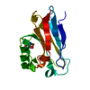
| ||||||||||||
|---|---|---|---|---|---|---|---|---|---|---|---|---|---|
| 1 |
| ||||||||||||
| Unit cell |
|
-Components
| #1: Protein | Mass: 14051.921 Da / Num. of mol.: 1 / Fragment: UNP residues 21-148 / Mutation: G116F Source method: isolated from a genetically manipulated source Source: (gene. exp.) Pseudomonas aeruginosa (bacteria) / Gene: azu, PA4922 / Production host: | ||||||||
|---|---|---|---|---|---|---|---|---|---|
| #2: Chemical |   Mass: 63.546 Da / Num. of mol.: 2 / Source method: obtained synthetically / Formula: Cu / Feature type: SUBJECT OF INVESTIGATION Mass: 63.546 Da / Num. of mol.: 2 / Source method: obtained synthetically / Formula: Cu / Feature type: SUBJECT OF INVESTIGATION#3: Chemical | ChemComp-TRS / |   Mass: 122.143 Da / Num. of mol.: 1 / Source method: obtained synthetically / Formula: C4H12NO3 / Comment: pH buffer*YM Mass: 122.143 Da / Num. of mol.: 1 / Source method: obtained synthetically / Formula: C4H12NO3 / Comment: pH buffer*YM#4: Water | ChemComp-HOH / |  Mass: 18.015 Da / Num. of mol.: 28 / Source method: isolated from a natural source / Formula: H2O Mass: 18.015 Da / Num. of mol.: 28 / Source method: isolated from a natural source / Formula: H2OHas ligand of interest | Y | Has protein modification | Y | |
-Experimental details
-Experiment
| Experiment | Method: X-RAY DIFFRACTION / Number of used crystals: 1 |
|---|
- Sample preparation
| Crystal | Density Matthews: 3.25 Å3/Da / Density % sol: 62.19 % |
|---|---|
| Crystal grow | Temperature: 277 K / Method: vapor diffusion, hanging drop / pH: 8 Details: 3 uL 1.4 mM in 100 mM sodium acetate, pH 5.6 + 1 uL reservoir buffer (20% PEG4000, 100 mM Tris-HCl, pH 8.0, 10 mM copper(II) sulfate, 100 mM lithium nitrate) against 250 uL reservoir buffer |
-Data collection
| Diffraction | Mean temperature: 100 K / Serial crystal experiment: N |
|---|---|
| Diffraction source | Source: SYNCHROTRON / Site: APS / Beamline: 23-ID-D / Wavelength: 1.03 Å |
| Detector | Type: DECTRIS PILATUS3 6M / Detector: PIXEL / Date: Sep 18, 2021 |
| Radiation | Monochromator: Si(111) / Protocol: SINGLE WAVELENGTH / Monochromatic (M) / Laue (L): M / Scattering type: x-ray |
| Radiation wavelength | Wavelength: 1.03 Å / Relative weight: 1 |
| Reflection | Resolution: 2.2→42.58 Å / Num. obs: 10424 / % possible obs: 99.6 % / Redundancy: 18 % / Biso Wilson estimate: 66.08 Å2 / CC1/2: 0.998 / Net I/σ(I): 20.78 |
| Reflection shell | Resolution: 2.2→2.279 Å / Num. unique obs: 1006 / CC1/2: 0.708 / % possible all: 100 |
- Processing
| Software |
| ||||||||||||||||||||||||||||||||||||||||||||||||||||||||
|---|---|---|---|---|---|---|---|---|---|---|---|---|---|---|---|---|---|---|---|---|---|---|---|---|---|---|---|---|---|---|---|---|---|---|---|---|---|---|---|---|---|---|---|---|---|---|---|---|---|---|---|---|---|---|---|---|---|
| Refinement | Method to determine structure: MOLECULAR REPLACEMENT Starting model: PDB entry 4AZU Resolution: 2.2→42.58 Å / SU ML: 0.3998 / Cross valid method: FREE R-VALUE / σ(F): 1.36 / Phase error: 29.7106 Stereochemistry target values: GeoStd + Monomer Library + CDL v1.2
| ||||||||||||||||||||||||||||||||||||||||||||||||||||||||
| Solvent computation | Shrinkage radii: 0.9 Å / VDW probe radii: 1.11 Å / Solvent model: FLAT BULK SOLVENT MODEL | ||||||||||||||||||||||||||||||||||||||||||||||||||||||||
| Displacement parameters | Biso mean: 73.93 Å2 | ||||||||||||||||||||||||||||||||||||||||||||||||||||||||
| Refinement step | Cycle: LAST / Resolution: 2.2→42.58 Å
| ||||||||||||||||||||||||||||||||||||||||||||||||||||||||
| Refine LS restraints |
| ||||||||||||||||||||||||||||||||||||||||||||||||||||||||
| LS refinement shell |
| ||||||||||||||||||||||||||||||||||||||||||||||||||||||||
| Refinement TLS params. | Method: refined / Origin x: 4.89749069655 Å / Origin y: -16.2824755597 Å / Origin z: 9.93376984892 Å
| ||||||||||||||||||||||||||||||||||||||||||||||||||||||||
| Refinement TLS group | Selection details: all |
